Topic Update

Recent Advance in Acute Lymphoblastic Leukaemia

Recent Advance in Acute Lymphoblastic Leukaemia


Volume 14, Issue 2, August 2019  (download full article in pdf)


Editorial note

In this topical update, Dr Albert Sin reviews recent advances in diagnosis and management of acute lymphoblastic leukaemia (ALL).  We welcome any feedback or suggestions. Please direct them to Dr Rock Leung (e-mail: leungyyr.ha.org.hk) of Education Committee, the Hong Kong College of Pathologists. Opinions expressed are those of the authors or named individuals and are not necessarily those of the Hong Kong College of Pathologists.


Dr. Albert Sin
Clinical Assistant Professor


Introduction

Acute lymphoblastic leukaemia (ALL) is an aggressive and highly fatal malignancy resulting from clonal mutations of lymphoid progenitor cells. The incidence of ALL is the most common in childhood and age after 50.18The prognosis of childhood ALL is good with long-term survival rate approaching 90% treated by intensive chemotherapy.12Although the incidence of ALL is less in adolescent, young adult as well as adult, the prognosis of ALL in those people is very poor, with only 30-40% of adult patients able to remit.18 According to the data from US database which registered all patients with diagnosed ALL from 2000 to 2007, the survival rate was 75% at 17 years old, 45% at 20 years old and 15% at 70 years old.14An increasing knowledge of disease biology of ALL transformed into insights for development of novel therapies to improve the treatment outcome of ALL.

One of the reasons of adverse prognosis in adolescent and young adult (AYA) as well as adult patients is that they commonly harbored poor-risk genetic aberrations while less patients carried favorable genetic lesion.14This could explain the sudden drop in survival from 17 years old to 20 years old. 


Ph-like ALL

Ph-like ALL is a newly identified genetic subgroup. The genetic profile of this subgroup of ALL is similar to that of Philadelphia chromosome positive (Ph-positive) ALL but without BCR-ABL1 fusion.17They have a higher frequency of IKZF1 deletion and mutation in genes of lymphoid transcription factors with poor survival.19The incidence of Ph-like ALL increases with ages and approaching 27% of cases of adult B-ALL.14

The nature of genetic aberration is heterogeneous. Despite its complexity, it can be simply classified into five subgroups: 1. CRLF2 rearrangement 2. Rearrangement of ABL-class gene 3. Rearrangement of JAK2 and EPOR 4. Aberrations leading to activation of JAK-STAT or MAPK pathway 5. Other rare kinase alterations.19The distribution of different types of genetic alterations are different among childhood high risk ALL, young adult and adult (Figure 1). CRLF2 rearrangement is the most common type of genetic alteration in Ph-like ALL. CRLF2 gene is responsible for producing lymphopoietin receptor and regulate the process of lymphopoiesis. Common mechanisms of CRLF2 rearrangement include 1. Translocation of CRLF2 gene into IGH gene 2. Fusion between CRLF2 gene and P2RY8 gene. 3. Point mutation F232C at CRLF2 gene. Nearly 50% of CRLF2 rearranged Ph-like ALL have concomitant JAK mutations.19


Figure 1

Figure 1


Diagnosis of Ph-like ALL


Genetic profiling is the gold standard for the diagnosis of Ph-like ALL. However, it is difficult to implement in routine diagnostic laboratory. 

Cytogenetics analysis is a standard test for all cases of ALL which allows a global assessment of chromosomal abnormalities. Some of the recurrent genetic abnormalities, for example t(9;22), hyperdiploidy/hyperdiploidy,   rearrangement involving 11q23, etc, can be detected. However, most of the Ph-like ALL genetic alterations are cryptic, e.g. interstitial deletion of CRLF2, ETV6-RUNX1 fusion, etc and thus they cannot be detected by conventional cytogenetics.2

Fluorescent in-situ hybridization (FISH) can be utilized to detect Ph-like ALL genetic abnormalities. Breakapart probes targeting genes most frequently genes including ABL1, ABL2, PDGFRB, JAK2, CRLF2, and P2RY8 are currently available. Although the positive result upon FISH study needs additional fusion probe for confirmation, it provides a readily available and useful diagnostic tool for establishing the diagnosis of Ph-like ALL. However, some of the important Ph-like ALL genetic rearrangement including intrachromosomal inversions (e.g., inv(9) resulting in PAX5-JAK2 fusion), intra-chromosomal deletions (e.g., del(X)(p22p22)/del(Y)(p11p11) resulting in P2RY8-CRLF2 fusion) are undetectable by FISH technique.2Targeted sequencing by NGS platform is an evolving technique for diagnosis.16

Recently, antibody against CRLF2 is available for flow cytometry study. The expression of CRLF2 as detected by multiparametric flow cytometry is correlated with genetic testing for CRLF2 rearrangement.15This provides a rapid tool for identifying potential cases of Ph-like ALL before the result of genetic tests is available. 

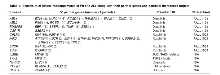
Most of the genetic alterations of Ph-like ALL are targetable kinase lesions, which could be treated by tailored kinase inhibitor therapy (Table 1).19This approach of therapy is current undergoing extensive preclinical studies.9


Table 1


Early T-cell precursor ALL (ETP-ALL)


ETP-ALL is recently characterized subtype of T-ALL. It constitutes around 12% of childhood ALL and 7.4% of adult ALL.11Genetic profiling showed ETP cells are similar to that of haemopoietic stem cells and myeloid progenitor cells.4This subgroup of ALL is characterized by the unique immunophenotype: cytoplasmic CD3+, surface CD3-, CD1a-, CD2-, CD5 dim (<75% positive), CD7 and positive for one or more stem cell and/or myeloid markers including HLA-DR, CD13, CD33, CD34, or CD117.5

While activating mutation of NOTCH1 is a common mutation found in ALL and it account for 50% of cases of childhood ALL, this mutation is less common in ETP-ALL.3ETP-ALL commonly have mutations in FLT3, DNMT3A, IDH1, IDH2, etc.11

ETP-ALL carries a poor prognosis with inferior overall survival when treated with standard chemotherapy regimen comparing with other subtypes of T-ALL.11This subgroup of T-ALL represented a distinct subtype with unique genetic profile and poor prognosis. 


MRD in adult ALL


Minimal residual disease (MRD) describe the very low level of disease burden which cannot be detected by morphology. Measurement of MRD not only pick up a submicroscopic level of disease but also can monitor the disease kinetic during the treatment process of haematological malignancies.10

The following techniques can be used to detect MRD: 

  1. Multiparametric flow cytometry to detect leukaemia-associated immunophenotype (LAIP)
    By using a 4-color or 6-color panel of antibodies, we can identify LAIP in 90% of ALL caes.10Flow cytometry is a quick method and the result of MRD can be generated in a short period of time for clinical decision. The sensitivity of MRD detection by this method is 0.01%. However, in order to define the positive MRD, we need 10-40 cluster of cells and thus higher number of cells are required for assessment which may be difficult for reassessment samples after intensive chemotherapy.7In addition, antigenic shift is commonly occurred in leukaemic cells and normal cells during the therapy. The use of monoclonal antibodies, e.g anti-CD19, anti-CD22 for treatment of ALL will affect the gating strategy used to identify the leukaemic cells.10

  2. Detecting leukaemia-specific fusion transcript by PCR technique
    Quantitative reverse-transcriptase PCR can be employed to detect the amount of leukaemia-specific fusion transcript. The sensitivity is higher compared with flow cytometry (10-4to 10-6).7The test is relatively easy to be performed in standardized diagnostic laboratory. However, only 30-40% of cases of ALL carry leukaemia-specific fusion transcript and thus limited the eligibility of MRD detection by this method. Moreover, the interpretation is challenging for RNA-based test in those cases will poor RNA quality.

  3. Quantitative PCR for immunoglobulin (IG)-T cell receptor (TCR) gene targets
    Quantitative PCR is employed to detect the specific sequence of rearranged IG gene or TCR gene in the sample. The sensitivity of this method is 10-4to 10-5and this method can be applied to all cases of ALL. However, this method of MRD detection requires prior characterization of IG or TCR gene rearrangement by sequencing and designs patient-specific primers for each case for subsequent MRD detection. Extensive standardization and experience are needed for the laboratory to set up this test, which limit the use of this method of MRD detection in diagnostic laboratory. Moreover, the clonal evolution in leukaemic blasts during treatment can make the original rearranged sequence to be lost and thus generate a false negative result. Also, the non-specific primer annealing occurs during the process of marrow regenerative may yield false positive result for the test.7


Application of MRD in treatment of adult ALL


MRD-guided therapy had been gained extensive experience in childhood ALL.8The study group of German Multicenter Study Group for Adult ALL (GMALL) had conducted largest study for the role of MRD in adult Ph-negative ALL. They showed that molecular remission is the only parameters significantly affect the remission duration and survival.6Patients with positive MRD after induction therapy achieved better overall survival after receiving haemopoietic stem cell transplant. Early achievement of MRD negativity after induction chemotherapy is associated with good outcome for adult ALL.10Study showed that MRD level correlates with post-transplant outcome.13Another group found that haemopoietic stem cell transplant benefits the patients with positive MRD at week 6.1These findings may prompt reconsideration of the indications of haemopoietic stem cell transplant for adult patients with ALL, especially those patients achieve MRD negativity after treatment. 


Concluding landmark


The prognosis of acute lymphoblastic leukaemia in young adolescent and adult is poor. The recent discovery of new subtype of acute lymphoblastic leukaemia with characterization of genetic lesions make a breakthrough of understanding of disease biology. Precise disease prognostication can be made. Targeted therapies are being developed for treating those patients. Clinical trials are conducting for evaluating the targeted therapies in those new subtypes of acute lymphoblastic leukaemia. Moreover, the application of MRD monitoring and MRD-adapted therapy in adult ALL can further stratified the patients and select the appropriate candidates of haemopoietic stem cell transplant in order to reduce transplant-related mortality and morbidity. The advances in understanding of molecular mechanism and disease biology of ALL help to improve the risk stratification, rapid development of targeted therapies and hopefully improve the prognosis in young adolescent and adult patients. 


Reference


  1. Beldjord K., Chevret S., Asnafi V., Huguet F., Boulland ML., Leguay T., Thomas X., Cayuela JM., Grardel N., Chalandon Y., Boissel N., Schaefer B., Delabesse E., Cavé H., Chevallier P., Buzyn A., Fest T., Reman O., Vernant JP., Lhéritier V., Béné MC., Lafage M., Macintyre E., Ifrah N., Dombret H; Group for Research on Adult Acute Lymphoblastic Leukemia (GRAALL). (2014) Oncogenetics and minimal residual disease are independent outcome predictors in adult patients with acute lymphoblastic leukemia. Blood, 2014,12;123(24):3739-49.
  2. Bradford J. Siegele., Valentina Nardi. (2018). Laboratory testing in BCR-ABL1-like (Philadelphia-like) B-lymphoblastic leukemia/lymphoma. Am J Hematol, 2018(93):971–977.
  3. Chao Gao., Shu-Guang Liu., Rui-Dong Zhang., Wei-Jing Li., Xiao-Xi Zhao., Lei Cui., Min-Yuan Wu., Hu-Yong Zheng and Zhi-Gang Li. (2014) NOTCH1 mutations are associated with favourable long-term prognosis in paediatric T-cell acute lymphoblastic leukaemia: a retrospective study of patients treated on BCH-2003 and CCLG-2008 protocol in China. Br J Haematol, 2014, 166(2):221-8. doi: 10.1111/bjh.12866
  4. Elaine Coustan-Smith., Charles G Mullighan., Mihaela Onciu., Frederick G Behm., Susana C Raimondi., Deqing Pei., Cheng Cheng., Xiaoping Su., Jeff rey E Rubnitz., Giuseppe Basso., Andrea Biondi., Ching-Hon Pui., James R Downing., Dario Campana. (2009) Early T-cell precursor leukaemia: a subtype of very high-risk acute lymphoblastic leukaemia.Lancet Oncol, 2009(10):147–56
  5. Elizabeth A. Raetz and David T. Teachey. (2016) T-cell acute lymphoblastic leukemia. Am Soc Haemaetolo Educ Program, 2016(1), 580-588
  6. Gökbuget N., Kneba M., Raff T., Trautmann H., Bartram CR., Arnold R., Fietkau R., Freund M., Ganser A., Ludwig WD., Maschmeyer G., Rieder H., Schwartz S., Serve H., Thiel E., Brüggemann M., Hoelzer D; German Multicenter Study Group for Adult Acute Lymphoblastic Leukemia. (2012) Adult patients with acute lymphoblastic leukemia and molecular failure display a poor prognosis and are candidates for stem cell transplantation and targeted therapies. Blood, 2012,120(9):1868-76.
  7. Jacques J. M. van Dongen., Vincent H. J. van der Velden., Monika Br¨uggemann and Alberto Orfao. (2015) Minimal residual disease diagnostics in acute lymphoblastic leukemia: need for sensitive, fast, and standardized technologies.Blood, 2015;125(26):3996-4009
  8. Marianne Ifversen., Dominik Turkiewicz., Hanne V. Marquart., Jacek Winiarski., Jochen Buechner., Karin Mellgren., Johan Arvidson., Jelena Rascon., Lenne-Triin Ko¨rgvee., Hans O. Madsen., Jonas Abrahamsson., Bendik Lund., Olafur G. Jonsson., Carsten Heilmann., Mats Heyman., Kjeld Schmiegelow., Kim Vettenranta. (2019) Low burden of minimal residual disease prior to transplantation in children with very high risk acute lymphoblastic leukaemia: The NOPHO ALL2008 experience. British Journal of Haematology, 2019(184): 982–993
  9. Maude SL, Tasian SK, Vincent T, et al. (2012) Targeting JAK1/2 and mTOR in murine xenograft models of Ph-like acute lymphoblastic leukemia. Blood, 2012;120(17):3510-3518.
  10. Monika Bru¨ ggemann and Michaela Kotrova. (2017) Minimal residual disease in adult ALL: technical aspects and implications for correct clinical interpretation. Hematology Am Soc Hematol Educ Program.2017(1):13-21. doi: 10.1182/asheducation-2017.1.13.
  11. Nitin Jain., Audrey V. Lamb., Susan O’Brien., Farhad Ravandi., Marina Konopleva., Elias Jabbour., Zhuang Zuo., Jeffrey Jorgensen., Pei Lin., Sherry Pierce., Deborah Thomas., Michael Rytting., Gautam Borthakur., Tapan Kadia., Jorge Cortes., Hagop M. Kantarjian., Joseph D. Khoury. (2016) Early T-cell precursor acute lymphoblastic leukemia/lymphoma (ETP-ALL/LBL) in adolescents and adults: a high-risk subtype. Blood, 2016;127(15):1863-1869
  12. Paul, S., Kantarjian, H., & Jabbour, E. J. (2016). Adult Acute Lymphoblastic Leukemia. Mayo Clin Proc, 91(11), 1645-1666. doi:10.1016/j.mayocp.2016.09.010
  13. Renato Bassan., Monika Brüggemann., Hoihen Radcliffe., Elizabeth Hartfield., Georg Kreuzbauer., Sally Wetten. (2019) A Systematic Literature Review And Meta-Analysis Of Minimal Residual Disease As A Prognostic Indicator In Adult B-Cell Acute Lymphoblastic Leukemia. Haematologica,2019 Mar 19. pii: haematol.2018.201053. doi: 0.3324/haematol.2018.201053.
  14. Roberts KG. (2018) Genetics and prognosis of ALL in children vs adults. Hematology Am Soc Hematol Educ Program. 2018(1):137-145. doi: 10.1182/asheducation-2018.1.137.
  15. Sergej Konoplev., Xinyan Lu., Marina Konopleva., Nitin Jain., Juan Ouyang., Maitrayee Goswami., Kathryn G. Roberts., Marc Valentine., Charles G. Mullighan., Carlos Bueso-Ramos., Patrick A. Zweidler-McKay., Jeffrey L. Jorgensen and Sa A. Wang. (2017) CRLF2-Positive B-Cell Acute Lymphoblastic Leukemia in Adult Patients: A Single-Institution Experience. Am J Clin Pathol, 2017(147):357-363
  16. Stadt UZ., Escherich G., Indenbirken D., Alawi M., Adao M., Horstmann MA. (2016) Rapid Capture Next-Generation Sequencing in Clinical Diagnostics of Kinase Pathway Aberrations in B-Cell Precursor ALL. Pediatr Blood Cancer, 2016;63(7):1283-6. doi: 10.1002/pbc.25975
  17. Tasian, S. K., Loh, M. L., & Hunger, S. P. (2017). Philadelphia chromosome-like acute lymphoblastic leukemia. Blood, 130(19), 2064-2072. doi:10.1182/blood-2017-06-743252
  18. Terwilliger, T., & Abdul-Hay, M. (2017). Acute lymphoblastic leukemia: a comprehensive review and 2017 update. Blood Cancer J, 7(6), e577. doi:10.1038/bcj.2017.53
  19. Thai Hoa Tran and Mignon L. Loh. (2016). Ph-like acute lymphoblastic leukaemia. Haematology Am Soc Haemaetolo Educ Program, 2016(1), 561-566.
Single Tags: 

Biochemical genetics in the expanded newborn screening era

topical_update_v14i1

Biochemical genetics in the expanded newborn screening era


Volume 14, Issue 1 January 2019  (download full article in pdf)


Editorial note

In this topical update, Dr. Calvin Chong reviews and updates on the technological development of biochemical genetics for the three major classes of metabolic disorders, namely aminoacidopathies, organic acidurias and fatty acid oxidation defects. These conditions have increasing local awareness, in particular with the introduction of universal expanded newborn screening. With a rising clinical demand for confirmatory tests in biochemical genetics, the ways to achieve better analytical quality and capacity were discussed. We welcome any feedback or suggestions. Please direct them to Dr. Sammy Chen (email: chenpls@ha.org.hk) of Education Committee, the Hong Kong College of Pathologists. Opinions expressed are those of the authors or named individuals, and are not necessarily those of the Hong Kong College of Pathologists.

Fig 0

Dr. Calvin Yeow-Kuan Chong
Department of Pathology, Princess Margaret Hospital


Introduction


Biochemical genetics refers to the diagnosis of genetic disorders with biochemical markers. Sir Archibald Garrod first described the biochemical features of alkaptonuria in 19021, and is often named as the founding father of biochemical genetics. Throughout the past century, the practice of biochemical genetics has evolved from spot chemical tests towards the use of chromatography and mass spectrometry2,3. The recent implementation of the pilot study of expanded newborn screening means, on one hand, disorders are diagnosed earlier, and patients with such conditions would fare better, and on the other hand, this represents an increase in the use of confirmatory tests in biochemical genetics which is most acutely felt at the major chemical pathology laboratories.

The screening panel of the government-initiated pilot study included 8 aminoacidopathies, 7 organic acidurias, 6 fatty acid oxidation defects, and 3 other disorders4. Apart from the three disorders which required separate measurements, all three other major classes of disorders (viz. the aminoacidopathies, organic acidurias, and fatty acid oxidation defects) are screened by the use of tandem mass spectrometric measurement of succinylacetone, amino acids and acylcarnitines, all done within a period of around two minutes.

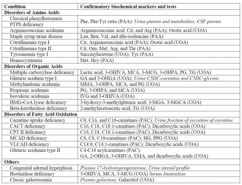
Table 1 lists the confirmatory markers and tests for the screened conditions 5,6: as can be seen, plasma amino acids, urine organic acids, and plasma acylcarnitines represents the main bulk work as confirmatory tests for these conditions. The present article aims therefore to explore the contemporary techniques available for these three major confirmatory tests in biochemical genetics and attempts to identify the approaches a laboratory may seek in order to cater for the increased demands.

Fig t

Table 1. Confirmatory markers and tests for conditions covered in the government-initiated pilot study of expanded newborn screening. Specialized tests not discussed in the present article are italicized.

Amino acids


Quantitative analysis of amino acids in plasma is the first-line confirmatory test for most aminoacidopathies. Amino acids are characterized by the presence of primary amine and carboxylic acid groups in one molecule, though some imino acids, namely proline and hydroxyproline, containing an imino (a functional group containing carbon-nitrogen double bond) as well as a carboxylic acid groups are also considered under the umbrella term amino acids in medical parlance and this use of terminology is adopted in the present article.

The analytical difficulties for amino acids are obvious when one examine their chemical structures: they are amphoteric, very small (glycine has a molecular mass of 75.067 g/mol), and most of them do not possess conjugated double bonds which gives absorbance in the ultraviolet spectra7. The first and second characteristics make separation difficult when reverse phase chromatography is used, whereas the last two give rise to problems in mass spectrometric detection and ultraviolet detection respectively. There are three major methods commonly used in clinical laboratories: amino acid analysers, high performance liquid chromatography (HPLC) with pre-column derivatization, and liquid chromatography-tandem mass spectrometry (LC-MS/MS). Irrespective of the methods employed, protein precipitation is necessary before analysis.

Amino acid analysers

Amino acid analysers (AAAs) are standalone machines that employ cation-exchange chromatography using a lithium buffer system, with post-column derivatization with ninhydrin (at some 120-135 degree Celsius) and monitoring at two wavelengths8. It is considered the gold standard method for amino acid analysis. This method overcomes the difficulty in separation of amino acids by utilizing ion-exchange chromatography which relies on the charge of a molecule at a particular pH rather than the hydrophobicity and steric interactions of a molecule and that of detection by formation of purple complexes which absorb strongly at 570 nm for primary amino acids, and yellow complexes which absorb strongly at 440 nm for proline and hydroxyproline9,10. When compared with other methods, AAAs provides higher degree of automation and require less expertise from the laboratory; the major issues with AAAs are the lengthy analytical run (120 minutes is common), the cost of the analytical instruments and the proprietary nature of the reagents. Analytical interference in AAAs are rare but do occur with dipeptides, which occur in prolidase deficiency8,11, and aspartylglucosamine, which occur in aspartylglucosaminuria8, can be spotted by the 570/440 absorbance ratio.

HPLC and LC-MS/MS methods

HPLC coupled with pre-column derivatization is used in the author’s laboratory for PAA determination. The method relied on the pre-column (immediately before injection) derivatization of amino acids by orthophthalaldehyde (OPA) and 3-mercaptopropionic acid (MPA), followed by reversed phase chromatography and ultraviolet detection12-14. In the presence of a thiol reagent, OPA forms a fluorescent derivative with primary amino acids with peak absorbance/excitation at 340 nm and emission wavelength at 450 nm7,8. The major drawback of this method is the inability to detect proline (and hydroxyproline) as OPA does not react with imino acids.

A newer derivatization reagent, AccQ-Tag (6-aminoquinolyl-N-hydroxysuccinimidyl carbamate), which converts both primary and secondary amino acids into stable fluorescent derivatives, has been used with ultra-high performance liquid chromatography with ultraviolet detection15. This derivatization reagent has the advantages of covering both primary and secondary amino acids and as the derivatization products are stable, advanced autosamplers which can pipet reagent from one vial to another is not required16. Complete chemistry kit-sets that use this derivatization reagent is commercially available17.

Liquid chromatography coupled with tandem mass spectrometry is also used locally for PAA determination. As these methods are often based on reversed phase chromatography, despite the use of mass spectrometric detector, derivatization is still often employed7. A method based on the proprietary derivatization reagent AccQ-Tag has been recently described in the literature with a run-time of 6 minutes18. Isotopic internal standards are required for LC-MS/MS-based methods and it is important to note that the impracticality of having isotopic internal standards for each and every analyte would mean that the robustness of the assay is considerably weaker than optical-based methods19.

Compared with methods based on optical detection, LC-MS/MS methods had a shorter analytical runtime and better specificity. This is counterweighed by the higher capital and maintenance cost of acquiring an LC-MS/MS (and not to mention the cost of having a backup LC-MS/MS system), as well as the technical expertise necessary to operate and troubleshoot an LC-MS/MS.

Choice of method and the future

The question is different depending on the volume of testing as well as equipment availability. For laboratories that has existing HPLC-DAD equipment employing OPA derivatization there may be little incentive in adopting a new procedure: there is probably little competition for HPLC analyser time, and the inability to detect proline may not be a major issue as hyperprolinaemia type I is benign whereas hyperprolinaemia type II is classically diagnosed by the presence of N-(Pyrrole-2-carboxyl)-glycine in urine organic acids20.

On the other hand, for a laboratory seeking to provide amino acids analysis for the first time, the use of stable derivatization reagent such as AccQ-Tag mentioned above has the advantage of not requiring higher-end liquid chromatographs and the availability of commercial kits means that development time is reduced. For a laboratory anticipating a high workload, a dedicated liquid chromatograph with UV detection appears to be the simplest solution as it is robust and inexpensive. On the other hand, for laboratories with lower service demand, mass spectrometric detection may in fact be more feasible as the notion of spare LC-MS/MS capacity means that the major downside of LC-MS/MS detection, viz capital cost and technical expertise, are sunk cost to the laboratory.

Organic acids

Organic acidurias are diagnosed most commonly by urine organic acid analysis with gas chromatography-mass spectrometry (GC-MS)21, and this technique is used by many local hospitals. Though the term organic acids refers to organic compounds with a carboxylic acid group, a broader spectrum of compounds, such as uracil and xanthine, are detected in practice. Urine organic analysis is usually qualitative though quantitative analyses of some compounds (e.g. orotic acid, methylmalonic acid) are often offered.

GC-MS based urine organic acid analysis

For GC-MS analysis of urine organic acids, a creatinine-corrected amount of urine is subject to liquid-liquid extraction with ethyl acetate after acidification using hydrochloric acid, the organic extract is then dried and derivatized by N,O-bis-tri(methylsilyl)trifluoroacetamide (BSTFA) with 1% trimethylchlorosilane (TMCS)22. The resultant product is injected to the GC-MS operating in scan mode. For reliable detection of ketones, oximation with hydroxylamine hydrochloride can be performed prior to acidic extraction23.

An alternative way of performing urine organic acid analysis is to bypass the extraction step. As no acidic extraction step is done, a limited panel of amino acids, purine, pyrimidines, and mono- and di-saccharides can be detected in the same analytical run. This method has been in-use in the author’s laboratory in the past 2 decades24,25, and allows the detection of a much wider range of metabolites in urine. With no extraction, the chromatograms are extremely complex due to co-elution of analytes and therefore this approach requires expertise in post-analytical data processing to be viable as a routine method.

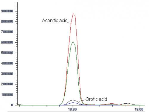
The interpretation of GC-MS data for urine organic acid analysis is commonly based on examination of the chromatogram (Figure 1), followed by a combination of peak integration in the total ion chromatogram followed by library searching and examination of extracted ion chromatogram at particular retention times for analytes which gives lower responses (Figure 2). The raw analyte response is then compared to locally-established age-specific reference intervals which allow clinical interpretation23.


Fig 1

Figure 1. Total ion chromatogram of a urine sample from a patient with malonic acidemia circulated in the ERNDIM qualitative organic acid program in 2016. The two abnormal peaks are indicated by asterisks: the first peak represents malonic acid (di-TMS derivative) and the second peak represents methylmalonic acid (di-TMS derivative).


Fig 2

Figure 2. Extracted ion chromatogram showing the quantifier ions (m/z 375, for aconitic acid in red; and m/z 254, for orotic acid, in black) and qualifier ions (m/z 285, for aconitic acid in green; and m/z 357, for orotic acid, in blue). They are co-eluting in many GC-MS based urine organic acid assays.


LC-MS based urine organic acid analysis

In the recent years, liquid chromatography-mass spectrometry-based methods have been published for analysis of urine organic acids. The benefit of liquid chromatography-mass spectrometry is clear: there is no need for the cumbersome derivatization step, and heat-labile analytes can be detected26,27. The major drawbacks of LC-MS based methods are the poorer separation of analytes and higher susceptibility towards ion suppression, as electrospray ionization is much less robust against matrix effects compared to electron ionization as used in GC-MS based methods28. The difficulty of developing an in-house LC-MS based method for urine organic acids lies in the procurement of a practically endless list of organic acid standards to establish the retention time and multiple reaction monitoring ratios. At the time of writing, there is at least one commercial kit that has been made available (Zivak Organic Acids LC-MS/MS analysis kit), but this commercial kit did not utilize isotopic internal standards even for critical analytes (such as hexanoylglycine and orotic acid) and one may wish to validate extensively the robustness of such assays against matrix effects.


Choice of method and the future

Out of the three assays discussed in the present article, urine organic acid analysis represents the most difficult of the three to establish in a laboratory. There is first the difficulty in establishing age-specific reference intervals, and then difficulty in acquiring a large number of chemical standards. A good starting point would be obtaining the bi-level quality controls for urine organic acid and special assays for urine from the ERNDIM network which consist of a number of critical analytes. As for choice of internal standards, while traditional choices such as tropic acid and pentadecanoic acid are often employed, deuterated internal standards covering critical analytes are much more available nowadays (e.g. methylmalonic acid-d3 and orotic acid-1,3-15N2) and their addition may improve quantitation of these critical analytes, the former being commonly quantified and the latter being prone to analytical errors29.

For laboratories with existing GC-MS based urine organic acid assay, the question is probably whether to adopt LC-MS based solution. The belief that organic aciduria always results in sky-high level of abnormal metabolites in urine is flawed: the low-excretor phenotype of glutaric aciduria type I can serve as the classical example30. The difficulty may be mitigated somewhat if acylcarnitines and acylglycines are measured by the LC-MS based assays as glutarylcarnitine has been shown to be informative even for low-excretor GA I patients31. Overall, it remains to be seen whether LC-MS based organic acid analysis could stand alone replacing, rather than complementing, GC-MS based assays.


Free carnitine and acylcarnitines


Quantitative analysis of free carnitine and acylcarnitines in plasma represents the first-line confirmatory test for fatty acid oxidation disorders. Analysis of free carnitine with calculation of fraction of excretion can be helpful in the diagnosis of carnitine uptake defect, as is measurement of urine glutarylcarnitine for the diagnosis of glutaric aciduria type I as discussed above31. Carnitine is a quaternary ammonium compound with a carboxylic acid group, as well as a hydroxyl group with which acyl groups are attached to form acylcarnitines; as such, it forms zwitterions under physiological pH. Derivatization to esters by incubation with an alcohol (typically butanol) has been employed32 though locally underivatized analysis at acidic pH is commonly used33.

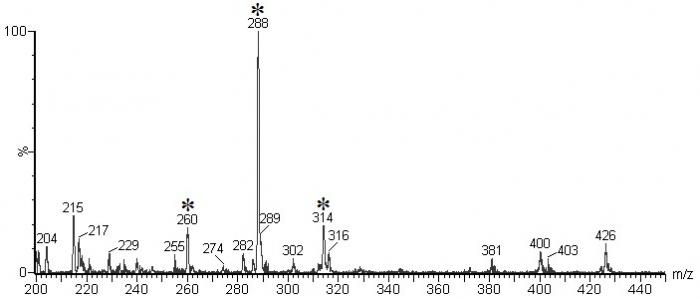
For underivatized analysis, plasma is first diluted with a mixture of isotopic internal standards, and acidified acetonitrile is slowly added to precipitate plasma proteins. The sample is vortexed, centrifuged, followed by evaporation of the supernatant to dryness and reconstituted for analysis by positive electrospray ionization-tandem mass spectrometry with or without liquid chromatography separation. Data can be collected in multiple reaction monitoring (MRM) mode, or in precursor ion scan with accumulation of ions, commonly known as multichannel acquisition (MCA) mode34 (Figure 3). The American College of Medical Genetics Guideline, published in 2008, suggested the use of precursor ion scan as it permitted the evaluation of the whole acylcarnitine profile, as well as the detection of drug artefacts, interfering compounds and assessment of derivatization35.

Fig 3

Figure 3. Cumulative precursor scan mass spectrum for a blood-spot sample distributed in the ERNDIM Qualitative Acylcarnitine Program in 2014. In this specimen, elevated signals at m/z ratio 260 (C6-carnitine), 288 (C8-carnitine), and 314 (C10:1-carnitine) can be seen. The pattern would be compatible with MCAD deficiency. (Mass spectra courtesy of Mr CK Lai, Chemical Pathology Laboratory, PMH)


Derivatization and chromatographic separation

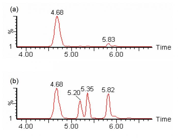
In the analysis of carnitine and acylcarnitines, butyl-ester derivatization enhances the formation of positively-charged ion by reacting with carboxylic groups, and causes mass separation of dicarboxylic-carnitines and hydroxy-acylcarnitines (e.g. C4DC-carnitine and C5OH-carnitine), which are isobaric when underivatized36. Derivatization is typically performed at highly acidic conditions (e.g. 3N hydrochloric acid at 65°C for 15 minutes)35. On the other hand, chromatographic separation allows the separate determination of individual isomeric constituents (e.g. C4DC-carnitines include succinylcarnitine and methylmalonylcarnitine; and C5OH-carnitines include 3-hydroxyisovalerylcarnitine and 2-methyl-3-hydroxybutyrylcarnitine) (Figure 4). With meticulous chromatographic separation, most biologically relevant isomeric species could be separately quantified37.


Choice of method and the future

The question for the laboratory is, first, whether to employ the derivatization procedure. The advantage of derivatization is the mass separation of hydroxylacylcarnitines and carnitine derivatives of dicarboxylic acids; the problems associated with derivatization are the partial hydrolysis of acylcarnitines because of the high temperature and strongly acidic condition employed33. The other considerations are whether to employ chromatography and if so, how extensive should it be: it would then be a delicate balance between through-put, diagnostic specificity, and analyser-time that is available. With the improvement of separation capability of liquid chromatographs and sensitivity of modern mass spectrometers, it is suggested that a short UHPLC program combined with both scheduled MRM and precursor ion scan function would be a good compromise.

Fig 4

Figure 4. SRM chromatogram of m/z 262>85 showing chromatographic separation of different species of isobaric (C4DC/C5OH) acylcarnitines (viz. succinylcarnitine at 4.68 minutes, two diastereomeric peaks of methylmalonylcarnitine at 5.2 and 5.35 minutes, and 3-hydroxyisovalerylcarnitine at 5.82 minutes) could be individually identified and quantified with liquid chromatography-tandem mass spectrometry without derivatization. (a) sample with normal amounts of succinylcarnitine; (b) sample with abnormal amounts of methylmalonylcarnitines (Chromatograms courtesy of Mr CK Lai, Chemical Pathology Laboratory, PMH)


Conclusions


The three assays discussed above represent the bulk of workload for most metabolic laboratories. The planned implementation of universal expanded newborn screening in Hong Kong means that the demand would increase, and the phenotype will be less defined, as tests are requested for patients who may not yet present with features of an inborn error and importantly, patients with only borderline elevation of analytes.

From a Bayesian point of view, this change in pre-test probability would mean that, if the analytical quality and interpretative capacity remains the same (which affect the likelihood ratio of positive results), the post-test probability would suffer from a negative impact. The quest for the metabolic bench of any major pathology laboratory is then to improve both throughput and quality of analysis at the same time, an impossible task, as the Duke of Norfolk wrote in 1538, “a man can not have his cake and eat his cake”38. The technology improvements as reviewed in the present article may aid in the analytical quality but the quest for improved interpretative capacity remains on the training and education of our present and coming generations of pathologists.


Reference

  1. Garrod, AE. The incidence of alkaptonuria: a study in chemical individuality. The Lancet. 1902 Dec 13;160(4137):1616–20.

  2. Millington DS, Kodo N, Norwood DL, Roe CR. Tandem mass spectrometry: a new method for acylcarnitine profiling with potential for neonatal screening for inborn errors of metabolism. Journal of inherited metabolic disease. 1990;13(3):321–4.

  3. Schulze A, Lindner M, Kohlmüller D, Olgemöller K, Mayatepek E, Hoffmann GF. Expanded newborn screening for inborn errors of metabolism by electrospray ionization-tandem mass spectrometry: results, outcome, and implications. Pediatrics. 2003;111(6):1399–406.

  4. Mak, CM. Newborn Screening: Past, Present and the Future. Topical Update, The Hong Kong College of Pathologists. 2016;11(2):1–10.

  5. Ozben T. Expanded newborn screening and confirmatory follow-up testing for inborn errors of metabolism detected by tandem mass spectrometry. Clinical Chemistry and Laboratory Medicine. 2012;51(1):157–176.

  6. Saudubray J-M, Berghe G van den, Walter JH. Inborn Metabolic Diseases. Springer-Verlag Berlin Heidelberg; 2012.

  7. Kaspar H, Dettmer K, Gronwald W, Oefner PJ. Advances in amino acid analysis. Anal Bioanal Chem. 2009 Jan;393(2):445–52.

  8. Duran M. Amino Acids. In: Blau N, Duran M, Gibson KM, editors. Laboratory Guide to the Methods in Biochemical Genetics [Internet]. Berlin, Heidelberg: Springer Berlin Heidelberg; 2008 [cited 2018 Nov 24]. p. 53–89. Available here.

  9. Friedman M, Williams LD. Stoichiometry of formation of Ruhemann’s purple in the ninhydrin reaction. Bioorganic Chemistry. 1974;3(3):267–80.

  10. Cunico RL, Schlabach T. Comparison of ninhydrin and o-phthalaldehyde post-column detection techniques for high-performance liquid chromatography of free amino acids. Journal of Chromatography A. 1983;266:461–70.

  11. Ferreira CR, Cusmano-Ozog K. Spurious Elevation of Multiple Urine Amino Acids by Ion-Exchange Chromatography in Patients with Prolidase Deficiency. JIMD Rep. 2016 Apr 12;31:45–9.

  12. Fürst P, Pollack L, Graser TA, Godel H, Stehle P. Appraisal of four pre-column derivatization methods for the high-performance liquid chromatographic determination of free amino acids in biological materials. Journal of Chromatography A. 1990;499:557–69.

  13. Schuster R. Determination of amino acids in biological, pharmaceutical, plant and food samples by automated precolumn derivatization and high-performance liquid chromatography. Journal of Chromatography B: Biomedical Sciences and Applications. 1988;431:271–84.

  14. Terrlink T, Van Leeuwen PA, Houdijk A. Plasma amino acids determined by liquid chromatography within 17 minutes. Clinical chemistry. 1994;40(2):245–9.

  15. Narayan SB, Ditewig-Meyers G, Graham KS, Scott R, Bennett MJ. Measurement of plasma amino acids by Ultraperformance® Liquid Chromatography. Clinical Chemistry and Laboratory Medicine. 2011;49(7):1177–1185.

  16. van Wandelen C, Cohen SA. Using quaternary high-performance liquid chromatography eluent systems for separating 6-aminoquinolyl-N-hydroxysuccinimidyl carbamate-derivatized amino acid mixtures. Journal of Chromatography A. 1997;763(1–2):11–22.

  17. Armenta JM, Cortes DF, Pisciotta JM, Shuman JL, Blakeslee K, Rasoloson D, et al. A sensitive and rapid method for amino acid quantitation in malaria biological samples using AccQ•Tag UPLC-ESI-MS/MS with multiple reaction monitoring. Anal Chem. 2010 Jan 15;82(2):548–58.

  18. Gray N, Zia R, King A, Patel VC, Wendon J, McPhail MJW, et al. High-Speed Quantitative UPLC-MS Analysis of Multiple Amines in Human Plasma and Serum via Precolumn Derivatization with 6-Aminoquinolyl-N-hydroxysuccinimidyl Carbamate: Application to Acetaminophen-Induced Liver Failure. Anal Chem. 2017 Feb 21;89(4):2478–87.

  19. Sharer JD, Biase ID, Matern D, Young S, Bennett MJ, Tolun AA. Laboratory analysis of amino acids, 2018 revision: a technical standard of the American College of Medical Genetics and Genomics (ACMG). Genetics in Medicine. 2018 Oct 19;1.

  20. Walker V, Mills GA. N-(pyrrole-2-carboxyl) glycine a diagnostic marker of hyperprolinaemia type II: mass spectra of trimethylsilyl derivatives. Clin Chim Acta. 2009 Jul;405(1–2):153–4.

  21. Christou C, Gika HG, Raikos N, Theodoridis G. GC-MS analysis of organic acids in human urine in clinical settings: a study of derivatization and other analytical parameters. Journal of Chromatography B. 2014;964:195–201.

  22. Rinaldo P. Organic Acids. In: Blau N, Duran M, Gibson KM, editors. Laboratory Guide to the Methods in Biochemical Genetics [Internet]. Berlin, Heidelberg: Springer Berlin Heidelberg; 2008 [cited 2018 Nov 30]. p. 137–69. Available here.

  23. Gallagher RC, Pollard L, Scott AI, Huguenin S, Goodman S, Sun Q, et al. Laboratory analysis of organic acids, 2018 update: a technical standard of the American College of Medical Genetics and Genomics (ACMG). Genet Med. 2018;20(7):683–91.

  24. Lee H-CH, Lai C-K, Yau K-CE, Siu T-S, Mak CM, Yuen Y-P, et al. Non-invasive urinary screening for aromatic l-amino acid decarboxylase deficiency in high-prevalence areas: A pilot study. Clinica Chimica Acta. 2012 Jan 18;413(1):126–30.

  25. Au KM, Lai CK, Yuen YP. Diagnosis of dihydropyrimidine dehydrogenase deficiency in a neonate with thymine-uraciluria. Hong Kong Medical Journal. 2003;9(2):130–3.

  26. Körver-Keularts IMLW, Wang P, Waterval HWAH, Kluijtmans LAJ, Wevers RA, Langhans C-D, et al. Fast and accurate quantitative organic acid analysis with LC-QTOF/MS facilitates screening of patients for inborn errors of metabolism. J Inherit Metab Dis. 2018 May 1;41(3):415–24.

  27. Yassine MM, Dabek-Zlotorzynska E. Application of ultrahigh-performance liquid chromatography–quadrupole time-of-flight mass spectrometry for the characterization of organic aerosol: Searching for naphthenic acids. Journal of Chromatography A. 2017 Aug 25;1512:22–33.

  28. Taylor PJ. Matrix effects: the Achilles heel of quantitative high-performance liquid chromatography–electrospray–tandem mass spectrometry. Clinical biochemistry. 2005;38(4):328–34.

  29. Scott C, Langhans C-D, Roux C. Qualitative Organic Acid. In: ERNDIM Workshop. Manchester, UK; 2017.

  30. Kölker S, Christensen E, Leonard JV, Greenberg CR, Boneh A, Burlina AB, et al. Diagnosis and management of glutaric aciduria type I–revised recommendations. Journal of inherited metabolic disease. 2011;34(3):677.

  31. Tortorelli S, Hahn SH, Cowan TM, Brewster TG, Rinaldo P, Matern D. The urinary excretion of glutarylcarnitine is an informative tool in the biochemical diagnosis of glutaric acidemia type I. Molecular Genetics and Metabolism. 2005 Feb 1;84(2):137–43.

  32. Dhillon KS, Bhandal AS, Aznar CP, Lorey FW, Neogi P. Improved tandem mass spectrometry (MS/MS) derivatized method for the detection of tyrosinemia type I, amino acids and acylcarnitine disorders using a single extraction process. Clinica Chimica Acta. 2011;412(11–12):873–9.

  33. Ho CS, Cheng BSS, Lam CWK. Rapid Liquid Chromatography–Electrospray Tandem Mass Spectrometry Method for Serum Free and Total Carnitine. Clinical Chemistry. 2003 Jul 1;49(7):1189–91.

  34. Matern D. Acylcarnitines, Including In Vitro Loading Tests. In: Blau N, Duran M, Gibson KM, editors. Laboratory Guide to the Methods in Biochemical Genetics [Internet]. Berlin, Heidelberg: Springer Berlin Heidelberg; 2008 [cited 2018 Nov 30]. p. 171–206. Available here.

  35. Rinaldo P, Cowan TM, Matern D. Acylcarnitine profile analysis. Genetics in Medicine. 2008 Feb;10(2):151–6.

  36. Vreken P, Van Lint AEM, Bootsma AH, Overmars H, Wanders RJA, Van Gennip AH. Quantitative plasma acylcarnitine analysis using electrospray tandem mass spectrometry for the diagnosis of organic acidaemias and fatty acid oxidation defects. Journal of inherited metabolic disease. 1999;22(3):302–6.

  37. Giesbertz P, Ecker J, Haag A, Spanier B, Daniel H. An LC-MS/MS method to quantify acylcarnitine species including isomeric and odd-numbered forms in plasma and tissues. Journal of lipid research. 2015;jlr. D061721.

  38. Letters and Papers, Foreign and Domestic, Henry VIII, Volume 13 Part 1, January-July 1538 [Internet]. 1892. Available here.


Abbreviations:
Plasma amino acids (PAA):
Arginine (Arg), Citrulline (Cit), Homocystine (Hcy), Isoleucine (Ileu), Leucine (Leu), Methionine (Met), Ornithine (Orn), Phenylalanine (Phe), Threonine (Thr), Tyrosine (Tyr), Valine (Val).

Urine organic acids (UOA):
ethylmalonic acid (EMA), glutaric acid (GA), 2-hydroxyglutaric acid (2-OHGA), 3-hydroxyglutaric acid (3-OHGA), 3-methylglutaric acid (3-MGA), 3-methylglutaconic acid (3-MGCA), hexanoylglycine (HG), 3-hydroxyisovaleric acid (3-OHIVA), Isovalerylglycine (IVG), 3-methylcrotonylglycine (3-MCG), Methylcitric acid (MCA), Methylmalonic acid (MMA), 3-hydroxypropionic acid (3-OHPA), propionylglycine (PG), phenylpropionylglycine (PPG), Tiglylglycine (TG).

Plasma acylcarnitines (PAC):
Free carnitine (C0).

Others:
6-pyruvoyl-tetrahydropterin synthase (PTPS), 3-hydroxy-3-methylglutaryl CoA (HMG-CoA), Carnitine-acylcarnitine translocase (CACT), Carnitine palmitoyltransferase II (CPT-II), Medium-chain acyl-CoA dehydrogenase (MCAD), Very long-chain acyl-coA dehydrogenase (VLCAD).
Single Tags: 

Antimicrobial resistance – a global health crisis

Antimicrobial resistance – a global health crisis


Volume 13, Issue 2 July 2018  (download full article in pdf)


Editorial note

With increasing prominence of the threat of antimicrobial resistance both internationally and locally, awareness and knowledge on the problem and prospects are essential, in order for rational application of control measures and monitoring of their effectiveness. In this issue of the Topical Update, Dr. Dominic Tsang and Dr. Christopher Lai present an updated overview of this important subject. We welcome any feedback or suggestion. Please direct them to Dr. Janice Lo (e-mail: janicelo@dh.gov.hk), Education Committee, The Hong Kong College of Pathologists. Opinions expressed are those of the authors or named individuals, and are not necessarily those of the Hong Kong College of Pathologists. 

Dr. Dominic TSANG and Dr. Christopher LAI
Consultant Microbiologist and Associate Consultant, Department of Pathology, Queen Elizabeth Hospital, Hong Kong 


End of modern medicine as we know it

The World Health Organization (WHO) described in 2001 antimicrobial resistance (AMR) as a global problem and an impending crisis. The apocalyptic term “Post-antibiotic era” was mentioned.1 The situation did not improve since. In fact, it deteriorated. In the United Kingdom (UK) government-commissioned “Review of Antimicrobial Resistance” by Lord Jim O’Neill, it was estimated that 700,000 people died from AMR infections in 2016 in the UK alone, and that drug-resistant infections could cause 10 million human deaths annually by 2050, costing the world up to $100 trillion.2 The Chief Medical Officer for England, Dame Professor Sally Davies, has predicted that unless tackled now, AMR could lead to the end of modern medicine as we know it. It could lead to routine operations and even childbirth becoming increasingly dangerous without the required antibiotics. In the UK, over 25,000 deaths a year are attributed to drug resistant infections.3 European Commission estimated the costs associated with AMR infection at €1.5 billion annually.4

The driving force behind emergence and dissemination of AMR is directly related to the use of antibiotics, i.e. the antibiotic selection pressure.5 It is recognized that non-human indiscriminate use of antibiotics in agriculture and animal husbandry to promote growth in animals and the consequential persistence of antibiotics in soil and aquatic environment select for AMR that could be disseminated widely.6 Human overuse nonetheless needs to be controlled by dedicated efforts on strengthening regulations on over-the- counter purchase of prescription only antibiotics, enhancing training in antibiotic prescriptions, monitoring compliance with antibiotic guideline and antibiotic stewardship programme (ASP).7


Concerted effort against AMR

Antibiotics on one hand is an agent required for life saving. But on the other, its usage and the resultant selective pressure have been recognized as the main drivers for AMR. Therefore, its use in human and non-human settings should be balanced against the risk of driving AMR. As such, antibiotic usage data has been linked to AMR surveillance data in the AMR containment strategy.8,9

There are already in place a few surveillance systems on AMR, covering healthy animals, diseased animals, food and humans in countries such as Canada, Denmark, Finland, Germany, Italy, Japan, USA, the Netherlands, Norway, France, and Sweden.10

The UK and China will establish the Global AMR Research Innovation Fund and encourage further investment from other governments and the private sector, helping to address AMR. The new fund will invite bids from industry, academia and other bodies. It aims to create international partnerships to build a global response and support new research to reduce the spread of antibiotic resistance.

The World Health Assembly in May 2015 endorsed a WHO global action plan to tackle antimicrobial resistance to ensure continuity of successful treatment and prevention of infectious diseases with effective and safe medicines.11 The plan sets out five strategic objectives including: to improve awareness and understanding of antimicrobial resistance; to strengthen knowledge through surveillance and research; to reduce the incidence of infection; to optimize the use of antimicrobial agents; and to develop the economic case for sustainable investment that takes account of the needs of all countries, and increase investment in new medicines, diagnostic tools, vaccines and other interventions. Under each of the objectives, specific actions were listed out for member states as well as international partners to implement.


Hong Kong Strategy against Multidrug resistant organisms (MDROs)

The Hong Kong Strategy and Action Plan on Antimicrobial Resistance 2017-202212 was released in July 2017 with the goals to develop a territory-wide network on surveillance on AMR and antimicrobial use, promote appropriate therapeutic use of antimicrobials in human and animals and to promote research on diagnostic and related interventions. Six key areas were targeted in the plan, including strengthening knowledge, optimizing antimicrobial use, reducing infections, improving awareness, promoting research and fostering partnerships among stakeholders.

In terms of AMR surveillance, the overall resistance profiles of MDROs have all along been closely monitored in public hospitals under the Hospital Authority in Hong Kong13 Among the concerned MDROs, Gram positive organisms include methicillin-resistant Staphylococcus aureus (MRSA) and vancomycin-resistant enterococcus (VRE). Gram negative organisms include extended spectrum beta-lactamase producing Enterobacteriaceae (ESBL-E) and the WHO top priority organisms of carbapenem-resistant Enterobacteriaceae (CRE), carbapenem- resistant Acinetobacter baumannii (CRAB), and carbapenem-resistant Pseudomonas aeruginosa (CRPA).14


Trends in antibiotic resistant organisms

MRSA: In Hong Kong, MRSA bloodstream infection was made a key performance indicator (KPI) to gauge the performance of infection control practices in all public hospitals.15 The corresponding MRSA bacteraemia rate was 0.18- 0.19 per 1,000 acute bed days when the monitoring began in 2008 and declined gradually to 0.144 per 1,000 acute bed days in 2017. At the same time, MRSA constituted 43.1% among S. aureus isolated from local HA hospitals.

VRE: The total number of VRE cases detected remains below 50 a month since 2015, after an upsurge and successful control in the Queen Elizabeth Hospital in 2013 when the peak number of cases detected in a month reached 300.16

CRAB/multi-drug resistant Acinetobacter baumannii (MDRA) is commonly associated with patients with prolonged hospital stay and who required ventilator-assisted ventilation. These patients are also usually put on multiple antibiotics for the treatment of underlying infections.17 Substantial environmental contamination with the resistant bacteria is frequent as a result of nursing care procedures, leading to explosive outbreaks which are difficult to abort unless attention is given to patient segregation and effective disinfection of instruments and environmental surfaces.18,19

ESBL-E began to emerge in late 1980s and peaked in UK in 2006 with 12% E. coli isolated from bacteraemic cases being ESBL positive. ESBL-E has been common locally, at 22% in 2017 compared to 24.3% since 2012. They are especially important as they commonly caused community-acquired as well as hospital-acquired infections. In fact, a local report showed 62% of imported chicken were found to be contaminated with ESBL-E.20 Community carriage rates for ESBL-E are high in East Mediterranean areas and South East Asia, from 30-60%.21,22 For treatment of ESBL-E infections, imipenem has been universally effective since its launch in 1987, and the same applies to meropenem, another carbapenem antibiotic which was launched in 1996.

CPE are members of the Enterobacteriaceae including E. coli and Klebsiella species which possess the gene on plasmids coding for the enzymes such as KPC, NDM and OXA-48 under the Ambler Classification on molecular class A, B or D respectively which enable them to inactivate carbapenem antibiotics and also other beta- lactams. CPE are usually multiply antibiotic resistant, therefore rendering limited options in treatment of their infections.23 The emergence and subsequent spread of CPE followed human movement closely as exemplified by the clustering of KPC-CPE in New York, North-West England, Israel and the spreading of NDM-CPE to Sweden by a returning patient from New Delhi, India.24-26 In Hong Kong, CPE cases were being increasingly detected, from 19 cases in 2011 to 473 cases in 2017. Active bacterial screening (ABS) of patients based on a set of consensus criteria in public hospitals including admission screening of all patients who have stayed in overseas hospitals in the past 12 months, suffering from antibiotic associated diarrhoea, staying in the same cubicle of a known CPE case, etc., helped to pick up an increasing number of CPE. Fortunately, most of the cases (90%) were asymptomatic carriers and did not require specific treatment. Of note, the number of confirmed CPE isolates in England in 2016 was more than 2,500 with KPC, NDM and OXA-48 predominant. Effective treatment options for CPE are few. The activities of ceftazidime and aztreonam with and without avibactam were tested against a large, contemporary, international collection of carbapenemase-producing Gram negative bacilli (CP-GNB) with diverse resistance mechanisms. Aztreonam-avibactam was active against all isolates except two NDM producers with elevated MICs of 8/4 and 16/4 mg/litre; ceftazidime- avibactam was active against all KPC-, IMI-, SME-, and most OXA-48 group-producing isolates (93%) but not metallo-β-lactamase producers. Among the older and contemporary antimicrobials, the most active were colistin, tigecycline, and fosfomycin, with overall susceptibilities of 88%, 79%, and 78%, respectively.27 Among local CPE isolates, the resistance rate of colistin is around 5.8%.


The “Find and Confine” Control strategy of AMR in HA hospitals

“Find”: This is the single most important control measures aiming to uncover the carriage state of any asymptomatic carrier, especially of VRE and CPE, who could shed the MDRO in the excreta. This is commonly done by ABS in the form of admission screening based on high risk factors such as hospitalization in recent months, a history of exposure to a known case, prolonged hospital stay (e.g. for 14 days or more), development of antibiotic associated diarrhoea etc. The yield of ABS at present is not high, ranging from 0.6% to 1.4% (average 1.0%) based on 2017 CPE ABS data in one of the hospitals, but ABS has undoubtedly provided a substantial impact in mitigating otherwise uninterrupted dissemination of VRE and CPE in the hospital setting. The availability of commercial agar media with the function to differentiate CPEs after overnight incubation has greatly facilitated ABS. Coupled with other rapid enzyme detection and PCR confirmation of CPE, which are also available commercially, the time to detection of CPE carriage in patients has been shortened to one to two days.

“Confine”: It is an important part of standard precautions (SP) plus contact precautions (CP) in the care of a known MDRO case. A room with en- suite is the preferred placement especially for those who suffer from diarrhoea and therefore at a higher risk of transmission of MDROs. This is widely practiced in developed countries. When en-suite rooms are not available, segregation of MDRO carriers by “cohorting” patients carrying the same MDRO is a pragmatic alternative.

Hand hygiene (HH): Healthcare workers (HCWs) strictly observing proper HH, e.g. WHO 5 moments, is undoubtedly the most important and effective measure in aborting the dissemination of MDROs in healthcare setting. The use of alcohol handrub in place of hand washing in situations where soiling is minimal has greatly improved HH compliance. However, HH is an action governed by human behavior which unfortunately also suffers from all factors that affect our behavior, such as physical fatigue, forgetfulness, motivation, persistence, peer influence, etc. For the same reason, the reliability of HH compliance monitoring by direct observation is also limited by the well-known “Hawthorn effect” in that, as humans, we tend to perform when we are aware of being observed.28 Further, in periods of high bed occupancy like influenza seasons, it is very hard to expect HCWs to fully comply with HH and as a consequence, these periods are more prone to cross transmission and outbreaks of MDROs. Apart from HCWs, patients and visitors are equally important in observing HH in order to avoid cross transmission. Similar moments of HH are being promoted for patients in local hospitals. In the long run, the development of a positive and motivated infection control culture and HH habit would not only maintain the cross transmission of MDROs at a low level but also mitigate the risk of other cross infections.

Environmental hygiene: The inanimate environment plays an important role inperpetuating the dissemination of MDROs in the hospital settings, especially VRE and MDRA.29,30 High-touch (frequently touched) surfaces such as patients’ privacy curtain, bed rails, door knobs, nursing trolleys, drip stands are often contaminated with the MDROs during outbreaks.31 The conventional cleansing and disinfection by using diluted sodium hypochlorite solution, although effective, is labour intensive. Also, to ensure the cleaning procedure is meticulously performed, it is commonly monitored by the use of a surrogate marker (e.g. UV-fluorescent marker) to ensure satisfactory performance.32 To circumvent such drawback, there are now plenty of effective new products on environmental disinfection, ranging from self- disinfecting surface coating sprays, hydrogen peroxide vapor, UV-C device, 2-in-1 disinfectant wipes to antimicrobial privacy curtains that could achieve effective decontamination with less labour. In a multi-centre study, 42.7% of standard hospital curtains were contaminated with MRSA and 42.3% with CRAB.33 The use of disposable antibacterial privacy curtains, e.g. nanoparticle silver or quaternary ammonium impregnated curtains, have been shown to prolong the time to contamination and reduce the bio-burden even after extended usage in acute care setting.34

Reducing overuse of antibiotics: It is imperative to maintain the use of antibiotic at the minimum essential level in order to prevent the emergence of resistance.34 Education and training at an early stage of the medical curriculum is critical in establishing the concept and skills in prudent antibiotic use. Guideline such as the local IMPACT guideline on antibiotic use is indispensable in providing the guidance on the right indications, choice, dose, route, and duration of antibiotic use. In addition, antibiotic stewardship program (ASP) has been widely practiced and proven to be effective in ensuring appropriate use of antibiotics.35 Locally, a multi- disciplinary team of clinical microbiologists, physicians, pharmacists and infection control nurses has been put in place in all HA hospitals to provide concurrent feedback on the use of targeted “big-gun” antibiotics. The percentage of appropriate use stays at above 80% in general and feedbacks on antibiotic use are welcomed in most of the cases.

Another very important aspect in the control of AMR is diagnostic support in infection.36 The rapid isolation and identification of an aetiological agent lends strong support in the continuation of antibiotic treatment, or its discontinuation in the absence of any evidence of infection. This is made possible with the introduction of molecular platforms such as 16S ribosomal RNA PCR and metagenomic studies.37

Surrogate biomarker of infection, in particular procalcitonin (PCT) which exhibits greater specificity than other proinflammatory markers such as C reactive protein (CRP) helps in identifying patients with sepsis and can be used for diagnosing infections, especially ventilator- associated pneumonia (VAP).38,39 The short half- life (25-30 hours in plasma) of PCT and its absence in healthy state make it the preferred biomarker for bacterial infections. PCT levels


The outlook of the challenges from AMR

The outcome in our battle against MDROs depends on how successful we are in preserving the efficacy of our existing antibiotics for treatment of infections and the result of our search for new antibiotics. Both require resources, efforts and dedication. While preserving existing effective antibiotics demands the aforementioned One Health approach, research breakthroughs arguably provide us the only hope in winning the battle against MDROs and to ensure effective treatment of infections for the continual practice of modern medicine.

Not long ago, researchers have identified from a soil sample a new cell wall inhibitor, teixobactin, from a previously unknown Gram-negative bacterium that lives in soil but which cannot be cultured in the laboratory using standard technique.40 The researchers used the “Ichip”, an isolation chamber, in which a soil sample is diluted with agar and a single bacterial cell in a chamber is then placed in soil where the bacteria could access to nutrients and growth factors. The teixobactin identified has excellent activity against Gram-positive pathogens including MRSA, Clostridium difficile, Bacillus anthracis and Mycobacterium tuberculosis. Another recent breakthrough was reported41 on accessing hidden natural products (NP) made by bacteria not by culturing but by sequencing, bioinformatics analysis and heterologous expression of biosynthetic gene clusters captured on DNA extracted from environmental samples. Such technique has led to the discovery of malacidins, a distinctive class of antibiotics which are active against MRSA infections without selection for resistance under the laboratory conditions. These discoveries are definitely good news given the great potential for more to be discovered by using these innovative technologies. Of course, there are still tests to be done before they could become clinically useful, but at least these discoveries through our human creative and innovative minds are holding promise in the battle against AMR. Meanwhile, we must not be distracted away from the momentum in tackling the rapidly deteriorating AMR situation.


Reference

  1. World Health Organization. WHO global strategy for containment of antimicrobial resistance [Internet]. Geneva: World Health Organization; 2001 [cited 2018 May].

  2. O’Neill J. Tackling Drug-Resistant Infections Globally: Final Report and Recommendations. London: Review on Antimicrobial Resistance; 2016 May 19 [cited 2018 May].

  3. McKie R. ‘Antibiotic apocalypse’: doctors sound alarm over drug resistance [Internet]. London: The Guardian; 2017 Oct 8 [cited 2018 May].

  4. Rodier G. European strategic action plan on antibiotic resistance 2011–2016. [Internet] Copenhagen: WHO Regional Office for Europe; 2011 Sep [cited 2018 May].

  5. Davies J, Davies D. Origins and evolution of antibiotic resistance. Microbiol Mol Biol Rev [Internet]. 2010 [cited 2010 Sep];74(3):417-33. In: Ovid MEDLINE(R) [Internet].

  6. Marshall BM, Levy SB. Food animals and antimicrobials: impacts on human health. Clin Microbiol Rev [Internet]. 2011 [cited 2011 Oct];24(4):718-33. In: Ovid MEDLINE(R) [Internet].

  7. Doron S, Davidson LE. Antimicrobial stewardship. Mayo Clin Proc [Internet]. 2011 [cited 2011 Nov];86(11):1113-23. In: Ovid MEDLINE(R) [Internet].

  8. World Health Organization. Global antimicrobial resistance surveillance system (GLASS) report: Early implementation 2016-2017 [Internet]. Geneva: World Health Organization; 2018 Jan 29 [cited 2018 May].

  9. Johnson I, Hansen A, Bi P. The challenges of implementing an integrated One Health surveillance system in Australia. Zoonoses Public Health [Internet]. 2018 [cited 2018 02];65(1):e229-e236. In: Ovid MEDLINE(R) In- Process & Other Non-Indexed Citations [Internet].

  10. Silley P, Simjee S, Schwarz S. Surveillance and monitoring of antimicrobial resistance and antibiotic consumption in humans and animals. Rev Sci Tech [Internet]. 2012 [cited 2012 Apr];31(1):105-20. In: Ovid MEDLINE(R) [Internet].

  11. World Health Organization. Global action plan on antimicrobial resistance [Internet]. Geneva: World Health Organization; 2015 [cited 2018 May]

  12. Centre for Health Protection. Hong Kong Strategy and Action Plan on Antimicrobial Resistance [Internet]. Hong Kong: Centre for Health Protection; 2018 May [cited 2018 May].

  13. Lai CK, Chuang WM, Kong MY, Siu HK, Tsang DN. Antimicrobial susceptibility in hospitals in Hong Kong: The current status 2009- 2011. J Glob Antimicrob Resist [Internet]. 2014 [cited 2014 Dec];2(4):225-231. In: Ovid MEDLINE(R) In-Process & Other Non-Indexed Citations [Internet].

  14. World Health Organization. WHO publishes list of bacteria for which new antibiotics are urgently needed [Internet]. Geneva: World Health Organization; 2017 Feb 27 [cited 2018 May].

  15. Leung PY. Making the Control of MRSA a Key Performance Indicator. Medical Diary [Internet]. 2011 Apr [cited 2018 May]; 16(4): 14.

  16. Lai CK, Wong SY, Lee SS, Siu HK, Chiu CY, Tsang DN, Ip MP, Hung CT. A hospital-wide screening programme to control an outbreak of vancomycin-resistant enterococci in a large tertiary hospital in Hong Kong. HONG KONG MED. J. [Internet]. 2017 [cited 2017 Apr];23(2):140-9. In: Ovid MEDLINE(R) [Internet].

  17. Huang H, Chen B, Liu G, Ran J, Lian X, Huang X, Wang N, Huang Z. A multi-center study on the risk factors of infection caused by multi- drug resistant Acinetobacter baumannii. BMC Infect Dis [Internet]. 2018 [cited 2018 01 05];18(1):11. In: Ovid MEDLINE(R) In-Process & Other Non-Indexed Citations [Internet]./a>

  18. Markogiannakis A, Fildisis G, Tsiplakou S, Ikonomidis A, Koutsoukou A, Pournaras S, Manolis EN, Baltopoulos G, Tsakris A. Cross- transmission of multidrug-resistant Acinetobacter baumannii clonal strains causing episodes of sepsis in a trauma intensive care unit. Infect Control Hosp Epidemiol [Internet]. 2008 [cited 2008 May];29(5):410-7. In: Ovid MEDLINE(R) [Internet]./a>

  19. Apisarnthanarak A, Pinitchai U, Thongphubeth K, Yuekyen C, Warren DK, Fraser VJ, Thammasat University Pandrug-Resistant Acinetobacter baumannii Control Group. A multifaceted intervention to reduce pandrug- resistant Acinetobacter baumannii colonization and infection in 3 intensive care units in a Thai tertiary care center: a 3-year study. Clin Infect Dis [Internet]. 2008 [cited 2008 Sep 15];47(6):760-7. In: Ovid MEDLINE(R) [Internet].

  20. Consumer Council. 測試 100 款生雞翼及全 雞 6 成驗出 ESBL 耐藥細菌 [Internet]. Hong Kong: Consumer Council; 2016 Dec 15 [cited 2018 May].

  21. Barreto Miranda I, Ignatius R, Pfuller R, Friedrich-Janicke B, Steiner F, Paland M, Dieckmann S, Schaufler K, Wieler LH, Guenther S, Mockenhaupt FP. High carriage rate of ESBL- producing Enterobacteriaceae at presentation and follow-up among travellers with gastrointestinal complaints returning from India and Southeast Asia. J Travel Med [Internet]. 2016 [cited 2016 Feb];23(2):tav024. In: Ovid MEDLINE(R) [Internet].

  22. Woerther PL, Andremont A, Kantele A. Travel-acquired ESBL-producing Enterobacteriaceae: impact of colonization at individual and community level. J Travel Med [Internet]. 2017 [cited 2017 Apr 01];24(suppl_1):S29-S34. In: Ovid MEDLINE(R) [Internet].

  23. Hrabak J, Chudackova E, Papagiannitsis CC. Detection of carbapenemases in Enterobacteriaceae: a challenge for diagnostic microbiological laboratories. Clin Microbiol Infect [Internet]. 2014 [cited 2014 Sep];20(9):839- 53. In: Ovid MEDLINE(R) [Internet].

  24. Woodford N, Tierno PM Jr, Young K, Tysall L, Palepou MF, Ward E, Painter RE, Suber DF, Shungu D, Silver LL, Inglima K, Kornblum J, Livermore DM. Outbreak of Klebsiella pneumoniae producing a new carbapenem- hydrolyzing class A beta-lactamase, KPC-3, in a New York Medical Center. Antimicrob Agents Chemother [Internet]. 2004 [cited 2004 Dec];48(12):4793-9. In: Ovid MEDLINE(R) [Internet].

  25. Samra Z, Ofir O, Lishtzinsky Y, Madar- Shapiro L, Bishara J. Outbreak of carbapenem- resistant Klebsiella pneumoniae producing KPC-3 in a tertiary medical centre in Israel. Int J Antimicrob Agents [Internet]. 2007 [cited 2007 Dec];30(6):525-9. In: Ovid MEDLINE(R) [Internet].

  26. Khong WX, Xia E, Marimuthu K, Xu W, Teo YY, Tan EL, Neo S, Krishnan PU, Ang BS, Lye DC, Chow AL, Ong RT, Ng OT. Local transmission and global dissemination of New Delhi Metallo-Beta-Lactamase (NDM): a whole genome analysis. BMC Genomics [Internet]. 2016 [cited 2016 06 13];17452. In: Ovid MEDLINE(R) [Internet].

  27. Vasoo S, Cunningham SA, Cole NC, Kohner PC, Menon SR, Krause KM, Harris KA, De PP, Koh TH, Patel R. In Vitro Activities of Ceftazidime-Avibactam, Aztreonam-Avibactam, and a Panel of Older and Contemporary Antimicrobial Agents against Carbapenemase-Producing Gram-Negative Bacilli. Antimicrob Agents Chemother [Internet]. 2015 [cited 2015 Dec];59(12):7842-6. In: Ovid MEDLINE(R) [Internet].

  28. Srigley JA, Furness CD, Baker GR, Gardam M. Quantification of the Hawthorne effect in hand hygiene compliance monitoring using an electronic monitoring system: a retrospective cohort study. BMJ Qual Saf [Internet]. 2014 [cited 2014 Dec];23(12):974-80. In: Ovid MEDLINE(R) [Internet].

  29. Chemaly RF, Simmons S, Dale C Jr, Ghantoji SS, Rodriguez M, Gubb J, Stachowiak J, Stibich M. The role of the healthcare environment in the spread of multidrug-resistant organisms: update on current best practices for containment. Ther. adv. infect. dis. [Internet]. 2014 [cited 2014 Jun];2(3-4):79-90. In: Ovid MEDLINE(R) In- Process & Other Non-Indexed Citations [Internet].

  30. Tan TY, Tan JS, Tay H, Chua GH, Ng LS, Syahidah N. Multidrug-resistant organisms in a routine ward environment: differential propensity for environmental dissemination and implications for infection control. J Med Microbiol [Internet]. 2013 [cited 2013 May];62(Pt 5):766-72. In: Ovid MEDLINE(R) [Internet].

  31. Dancer SJ. Controlling hospital-acquired infection: focus on the role of the environment and new technologies for decontamination. Clin Microbiol Rev [Internet]. 2014 [cited 2014 Oct];27(4):665-90. In: Ovid MEDLINE(R) [Internet].

  32. Ling ML, Apisarnthanarak A, Thu le TA, Villanueva V, Pandjaitan C, Yusof MY. APSIC Guidelines for environmental cleaning and decontamination. Antimicrob. resist. infect. control [Internet]. 2015 [cited 2015];458. In: Ovid MEDLINE(R) In-Process & Other Non-Indexed Citations [Internet].

  33. Shek K, Patidar R, Kohja Z, Liu S, Gawaziuk JP, Gawthrop M, Kumar A, Logsetty S. Rate of contamination of hospital privacy curtains on a burns and plastic surgery ward: a cross-sectional study. J Hosp Infect [Internet]. 2017 [cited 2017 May];96(1):54-58. In: Ovid MEDLINE(R) [Internet].

  34. Canton R, Morosini MI. Emergence and spread of antibiotic resistance following exposure to antibiotics. FEMS Microbiol Rev [Internet]. 2011 [cited 2011 Sep];35(5):977-91. In: Ovid MEDLINE(R) [Internet].

  35. Plachouras D, Hopkins S. Antimicrobial stewardship: we know it works; time to make sure it is in place everywhere. Cochrane Database Syst Rev [Internet]. 2017 [cited 2017 02 09];2ED000119. In: Ovid MEDLINE(R) Revisions [Internet].

  36. Center for Infectious Disease Research and Policy (CIDRAP). ASP Policy Update: Rapid Diagnostic Testing in Antimicrobial Stewardship [Internet]. Minneapolis: University of Minnesota; 2017 Oct [cited 2018 May].

  37. Bogaert D, van Belkum A. Antibiotic treatment and stewardship in the era of microbiota-oriented diagnostics. Eur J Clin Microbiol Infect Dis [Internet]. 2018 [cited 2018 05];37(5):795-798. In: Ovid MEDLINE(R) In- Process & Other Non-Indexed Citations [Internet].

  38. Stolz D, Smyrnios N, Eggimann P, Pargger H, Thakkar N, Siegemund M, Marsch S, Azzola A, Rakic J, Mueller B, Tamm M. Procalcitonin for reduced antibiotic exposure in ventilator- associated pneumonia: a randomised study. Eur Respir J [Internet]. 2009 [cited 2009 Dec];34(6):1364-75. In: Ovid MEDLINE(R) [Internet].

  39. Schuetz P, Wirz Y, Sager R, Christ-Crain M, Stolz D, Tamm M, Bouadma L, Luyt CE, Wolff M, Chastre J, Tubach F, Kristoffersen KB, Burkhardt O, Welte T, Schroeder S, Nobre V, Wei L, Bucher HC, Bhatnagar N, Annane D, Reinhart K, Branche A, Damas P, Nijsten M, de Lange DW, Deliberato RO, Lima SS, Maravic-Stojkovic V, Verduri A, Cao B, Shehabi Y, Beishuizen A, Jensen JS, Corti C, Van Oers JA, Falsey AR, de Jong E, Oliveira CF, Beghe B, Briel M, Mueller B. Procalcitonin to initiate or discontinue antibiotics in acute respiratory tract infections. Cochrane Database Syst Rev [Internet]. 2017 [cited 2017 10 12];10CD007498. In: Ovid MEDLINE(R) In- Process & Other Non-Indexed Citations [Internet].

  40. Ling LL, Schneider T, Peoples AJ, Spoering AL, Engels I, Conlon BP, Mueller A, Schaberle TF, Hughes DE, Epstein S, Jones M, Lazarides L, Steadman VA, Cohen DR, Felix CR, Fetterman KA, Millett WP, Nitti AG, Zullo AM, Chen C, Lewis K. A new antibiotic kills pathogens without detectable resistance. Nature [Internet]. 2015 [cited 2015 Jan 22];517(7535):455-9. In: Ovid MEDLINE(R) [Internet].

  41. Hover BM, Kim SH, Katz M, Charlop-Powers Z, Owen JG, Ternei MA, Maniko J, Estrela AB, Molina H, Park S, Perlin DS, Brady SF. Culture- independent discovery of the malacidins as calcium-dependent antibiotics with activity against multidrug-resistant Gram-positive pathogens. Nat. microbiol. [Internet]. 2018 [cited 2018 Apr];3(4):415-422. In: Ovid MEDLINE(R) In-Process & Other Non-Indexed Citations [Internet].

Single Tags: 

An overview of NMO Spectrum Disorder and the diagnostic utility of anti-NMO antibodies

An overview of NMO Spectrum Disorder and the diagnostic utility of anti-NMO antibodies


Volume 13, Issue 1 December 2017  (download full article in pdf)


Editorial note

NMOSD is an immune mediated demyelinating disease. Though its clinical presentation may overlap with multiple sclerosis, distinguishing these two entities is important in view of differences in treatment. Anti-NMO antibodies play a crucial role in the workup and diagnosis of NMOSD. In this review, Dr Elaine Au provided an overview of the NMOSD condition and the use of anti-NMO antibody assays. We welcome any feedback or suggestions. Please direct them to Dr Elaine Au (email: ayl436@ha.org.hk) of Education Committee, the Hong Kong College of Pathologists. Opinions expressed are those of the authors or named individuals, and are not necessarily those of the Hong Kong College of Pathologists. 

Dr Au Yuen Ling Elaine
Associate Consultant, Division of Clinical Immunology, Department of Pathology, Queen Mary Hospital 


NMO is an idiopathic immune mediated demyelinating disease that predominantly affects optic nerves and spinal cord. The prevalence range from 0.3 to 4.4 per 100000 people, with Asian and African-American more affected than Caucasian, where multiple sclerosis is more common in the white population (1-5). The condition has been named as Devic’s disease in the past (6), which described a monophasic disorder presenting with simultaneous bilateral optic neuritis and transverse myelitis. With the availability of specific serological marker, antibodies that targeted the water channel aquaporin-4 (AQP4), the clinical and neuroimaging spectrum of NMO disease is broadened. Instead of being a monophasic disorder, NMO antibodies positive patients with recurrent attacks are not uncommonly found. Moreover, the clinical presentations are more variable than the traditional Devic disease. There is no pathognomonic clinical feature of spectrum disorder (NMOSD), though certain clinical presentations are particularly suggestive of the disorder. These include simultaneously bilateral optic neuritis, complete spinal cord syndrome and area postrema clinical syndrome.


Multiple sclerosis is an important differential diagnosis of this condition in view of the overlapping clinical features of these two conditions. In NMOSD, optic neuritis tends to be more severe, more often with simultaneous bilateral involvement or sequential in rapid succession, compare to multiple sclerosis. Complete spinal cord syndrome, with longitudinally extensive transverse myelitis in MRI, is more suggestive of NMOSD than multiple sclerosis. Differentiating NMOSD from other demyelinating disease, i.e. multiple sclerosis is important since the treatment is different. Indeed, some multiple sclerosis therapies may aggravate NMO disorders (7-10).


Diagnostic Criteria

In 2006, the serological marker was first incorporated into the revised NMO diagnostic criteria. In 2007, NMOSD was introduced to include seropositive patients who do not follow the classical monophasic bilateral optic neuritis and transverse myelitis. Lately, the International Panel for NMO Diagnosis (IPND) has further revised the diagnostic criteria (11). For anti-NMO antibodies positive cases, presenting at least one core clinical characteristic of the disease is required for diagnosis. Core clinical characteristics include optic neuritis, acute myelitis, area postrema syndrome (episode of otherwise unexplained hiccups or nausea and vomiting), acute brainstem syndrome, narcolepsy or acute diencephalic syndrome with typical Magnetic Resonance Imaging (MRI) lesions and symptomatic cerebral syndrome with typical MRI lesions. On the other hand, more stringent clinical criteria, with additional neuroimaging findings, are required in seronegative patients to fulfill the diagnostic criteria (See appendix). 

For the workup of the disease, MRI, cerebral spinal fluid (CSF) exam and serological test for anti-NMO antibodies are important. In some patients, CSF pleocytosis, usually in the form of monocytosis and lymphocytosis, are present. Increased CSF protein levels are noted in 46-75% of cases (12, 13). Nevertheless, presence of CSF oligoclonal bands is uncommon in NMOSD, and at most transient, in contrast to the presence of persistent CSF oligoclonal bands in the case of multiple sclerosis. Finally, visual evoked potentials, somatosensory evoked potentials and brainstem acoustic evoked potentials examination may also be helpful in the workup.


Clinical course and prognosis

In NMOSD syndromes affecting regions other than optic nerve and spinal cord, not uncommonly patients will relapse with more classical involvement in subsequent attacks. Majority of NMOSD patients suffer from recurrent attacks (80- 90%), less frequently monophasic attack (10-20%) (14), while gradual progressive course with neurological deterioration is very rare (15). Relapses usually occur in clusters, but unpredictable intervals. In the Mayo Clinic series, the second relapse occurred within 1 year in 60% of cases, and within 3 years in 90% of cases (16). Repeated NMO attacks not uncommonly lead to accumulation of neurological impairment. NMO is associated with more adverse outcome than MS in general(14).

NMOSD has been shown to be frequently associated with other autoimmune disorders, including lupus, Sjogren’s syndrome, etc (17-19). On contrary, NMOSD is not common in rheumatic disease patients.


Anti-NMO antibodies

Among the different investigations, NMO antibodies test is central to the workup. Anti-NMO antibodies are pathogenic. The third extracellular loop of AQP4 is the major epitope for the anti- NMO antibodies. Biopsy and autopsy tissue obtained from seropositive patients demonstrate loss of AQP4 immunoreactivity. Perivascular complement activation in actively demyelinating lesions is also happened. In the central nervous system (CNS), AQP4 is expressed at the foot processes of astrocytes, near the basement membranes, in the optic nerve, in a subpopulation of ependymal cells, in hypothalamic nuclei and in the subfornical organ (20, 21). Truncated astrocyte processes or cell loss were found in demyelinating lesions (11). In rat models, passive transfer of the antibodies leaded to the development of disease (22, 23). These pathological findings distinguish NMOSD from multiple sclerosis.

The Anti-NMO antibodies can be detected by indirect immunofluorescence staining on tissue slide using mouse cerebellum tissue section, cell- based assays, radioimmunoprecipitation assays and enzyme-linked immunosorbent assays (ELISA). Overall, cell-based assay is preferred in view of better assay sensitivity and specificity compared to other methods. Ideally, confirmatory testing with one or more techniques is suggested (11), especially in cases with atypical presentation or borderline results are obtained.

In tissue based indirect immunofluorescence test, NMO antibodies positive case is characterized by the binding to structures adjacent to microvasculature, the Virchow-Robin spaces (VRS) and pia mater (Fig.1).


Fig 1

Fig. 1. Mouse cerebellum tissue section stained with anti-NMO antibodies.


This assay allows the detection of any coexisting anti-neuronal antibodies, which may be important as differential diagnosis and workup. However, the method is observer dependent and subjective. The interpretation of antibody staining may easily be affected by non-specific background staining on the tissue. In some rare occasions, antibodies other than anti-NMO may mimic the staining pattern and lead to false positive results. Moreover, indirect tissue based immunofluorescence test has relative low sensitivity (63-64%) (24-27).

Cell-based assay utilize cell lines such as human embryonic kidney 293 (HEK293) cells or Chinese hamster ovary (CHO) cells that have been transfected with AQP4 gene expression vector, so that expressing much higher level of antigen comparing to normal tissues. Cell lines from different units may use different ratio of the two isoform of AQP4: M1 and M23 in order to obtain optimal antigen presentation. Cell-based assay can be assessed by indirect immunofluorescence staining or flow cytometry. For indirect immunofluorescence cell- based assay, slide with fixed AQP-4 gene transfected cells and non- transfected cells growing on different biochips are placed side by side for comparison. Therefore, false positivity is minimized with the inclusion of control non-transfected cells. A higher expression of antigen in the transfected cells also enhance the assay sensitivity compared to tissue-based indirect immunofluorescence testing. (Fig.2)


Fig 1

Fig.2. HEK 293 cells transfected with AQP-4 gene expression vector and stained positive with anti- NMO antibodies.


Overall, cell-based assay is the recommended assay in view of good sensitivity and specificity (mean sensitivity 76.7% in a pooled analysis; 0.1% false positivity in a multiple sclerosis cohort) (24- 27). Commercial kits for indirect immunofluorescence cell-based assay are available, which facilitate the assay setup in service laboratories. Nevertheless, indirect immunofluorescence method is semi-quantitative and observer dependent.

Protein-based assays, like ELISA and radioimmunoprecipitation assays, in general, have lower sensitivity compared to cell based assays. In addition, ELISA, in particular at low titer, may yield nonspecific results. However, these assays provide quantitative results which may potentially be used for serial monitoring (24).

Though NMO antibody testing in serum is well- established, the diagnostic role of testing the antibody in CSF is controversial. Most of the cases reported in literature are diagnosed by serum test, though there have been rare cases reported that were CSF positive but serum test negative (28, 29). When studying paired CSF and serum samples with antibody indices calculated, intrathecal production of the NMO antibody is rare (24). NMO antibodies can be present in patients few years before and after the disease presentations. Lately, there is increasing evidence that the antibody titre may reflect disease activity. Elevated antibody levels at relapse and decrease in titre after immunosuppressant treatment has been reported in literature (30-34). Therefore, serial monitoring may possibly facilitate management and medication adjustment. However, there is no general threshold value for clinical relapse and the absolute level varies with individual patients. Rising level may not predict relapse in all cases. In addition, some methodologies, like indirect immunofluorescence test, only provide semi-quantitative results, and inter-run reproducibility is another issue. Other factors including the frequency of test necessary to achieve meaningful disease status monitoring and the cost involved are also important consideration. Therefore, it remains to be determined whether the marker should be serially monitored for treatment response and disease activity monitoring.


Treatment

The treatment for classical Devic’s disease presentation and relapsing NMOSD presentation is no different. High dose steroid is commonly employed as first- line of treatment in acute presentation. Plasma exchange may be considered in treatment refractory cases. Immunomodulatory treatment with interferon β, which is a treatment option in multiple sclerosis, may exacerbate NMOSD disorders. Therefore, differentiating between these two conditions is important. Options of steroid sparing immunosuppressants to consider in NMOSD include azathioprine, methotrexate, mycophenolate mofetil, rituximab, etc (14).


Conclusion

NMOSD is a rare but increasingly recognized condition, which present as an inflammatory and demyelinating autoimmune disorder affecting the central nervous system. With the availability of a serological marker, anti-NMO antibody, the diagnosis and differentiating from related conditions is facilitated. Timely diagnosis and treatment is important for the management of these patients.


Reference

  1. Kira J. Multiple sclerosis in the Japanese population. The Lancet Neurology. 2003;2(2):117- 27.

  2. abre P, Signate A, Olindo S, Merle H, Caparros-Lefebvre D, Bera O, et al. Role of return migration in the emergence of multiple sclerosis in the French West Indies. Brain : a journal of neurology. 2005;128(Pt 12):2899-910.

  3. Asgari N, Lillevang ST, Skejoe HP, Falah M, Stenager E, Kyvik KO. A population-based study of neuromyelitis optica in Caucasians. Neurology. 2011;76(18):1589-95.

  4. Cossburn M, Tackley G, Baker K, Ingram G, Burtonwood M, Malik G, et al. The prevalence of neuromyelitis optica in South East Wales. European journal of neurology. 2012;19(4):655-9.

  5. Mealy MA, Wingerchuk DM, Greenberg BM, Levy M. Epidemiology of neuromyelitis optica in the United States: a multicenter analysis. Archives of neurology. 2012;69(9):1176-80.

  6. Jarius S, Wildemann B. The history of neuromyelitis optica. Journal of neuroinflammation. 2013;10:8.

  7. Kimbrough DJ, Fujihara K, Jacob A, Lana- Peixoto MA, Leite MI, Levy M, et al. Treatment of Neuromyelitis Optica: Review and Recommendations. Multiple sclerosis and related disorders. 2012;1(4):180-7.

  8. Kleiter I, Hellwig K, Berthele A, Kumpfel T, Linker RA, Hartung HP, et al. Failure of natalizumab to prevent relapses in neuromyelitis optica. Archives of neurology. 2012;69(2):239-45.

  9. Min JH, Kim BJ, Lee KH. Development of extensive brain lesions following fingolimod (FTY720) treatment in a patient with neuromyelitis optica spectrum disorder. Multiple sclerosis. 2012;18(1):113-5.

  10. Papeix C, Vidal JS, de Seze J,Pierrot- Deseilligny C, Tourbah A, Stankoff B, et al. Immunosuppressive therapy is more effective than interferon in neuromyelitis optica. Multiple sclerosis. 2007;13(2):256-9.

  11. Wingerchuk DM, Banwell B, Bennett JL, Cabre P, Carroll W, Chitnis T, et al. International consensus diagnostic criteria for neuromyelitis optica spectrum disorders. Neurology. 2015;85(2):177-89.

  12. O'Riordan JI, Gallagher HL, Thompson AJ, Howard RS, Kingsley DP, Thompson EJ, et al. Clinical, CSF, and MRI findings in Devic's neuromyelitis optica. Journal of neurology, neurosurgery, and psychiatry. 1996;60(4):382-7.

  13. de Seze J, Stojkovic T, Ferriby D, Gauvrit JY, Montagne C, Mounier-Vehier F, et al. Devic's neuromyelitis optica: clinical, laboratory, MRI and outcome profile. Journal of the neurological sciences. 2002;197(1-2):57-61.

  14. Sellner J, Boggild M, Clanet M, Hintzen RQ, Illes Z, Montalban X, et al. EFNS guidelines on diagnosis and management of neuromyelitis optica. European journal of neurology. 2010;17(8):1019-32.

  15. Wingerchuk DM, Pittock SJ, Lucchinetti CF, Lennon VA, Weinshenker BG. A secondary progressive clinical course is uncommon in neuromyelitis optica. Neurology. 2007;68(8):603-5.

  16. Wingerchuk DM, Hogancamp WF, O'Brien PC, Weinshenker BG. The clinical course of neuromyelitis optica (Devic's syndrome). Neurology. 1999;53(5):1107-14.

  17. Jarius S, Jacobi C, de Seze J, Zephir H, Paul F, Franciotta D, et al. Frequency and syndrome specificity of antibodies to aquaporin-4 in neurological patients with rheumatic disorders. Multiple sclerosis. 2011;17(9):1067-73.

  18. Pittock SJ, Lennon VA, de Seze J, Vermersch P, Homburger HA, Wingerchuk DM, et al. Neuromyelitis optica and non organ-specific autoimmunity. Archives of neurology. 2008;65(1):78-83.

  19. Wandinger KP, Stangel M, Witte T, Venables P, Charles P, Jarius S, et al. Autoantibodies against aquaporin-4 in patients with neuropsychiatric systemic lupus erythematosus and primary Sjogren's syndrome. Arthritis and rheumatism. 2010;62(4):1198-200.

  20. Graber DJ, Levy M, Kerr D, Wade WF. Neuromyelitis optica pathogenesis and aquaporin 4. Journal of neuroinflammation. 2008;5:22.

  21. Tait MJ, Saadoun S, Bell BA, Papadopoulos MC. Water movements in the brain: role of aquaporins. Trends in neurosciences. 2008;31(1):37-43.

  22. Kinoshita M, Nakatsuji Y, Moriya M, Okuno T, Kumanogoh A, Nakano M, et al. Astrocytic necrosis is induced by anti-aquaporin-4 antibody- positive serum. Neuroreport. 2009;20(5):508-12.

  23. Kinoshita M, Nakatsuji Y, Kimura T, Moriya M, Takata K, Okuno T, et al. Neuromyelitis optica: Passive transfer to rats by human immunoglobulin. Biochemical and biophysical research communications. 2009;386(4):623-7.

  24. Jarius S, Wildemann B. Aquaporin-4 antibodies (NMO-IgG) as a serological marker of neuromyelitis optica: a critical review of the literature. Brain pathology. 2013;23(6):661-83.

  25. Waters PJ, McKeon A, Leite MI, Rajasekharan S, Lennon VA, Villalobos A, et al. Serologic diagnosis of NMO: a multicenter comparison of aquaporin-4-IgG assays. Neurology. 2012;78(9):665-71; discussion 9.

  26. Sato DK, Nakashima I, Takahashi T, Misu T, Waters P, Kuroda H, et al. Aquaporin-4 antibody- positive cases beyond current diagnostic criteria for NMO spectrum disorders. Neurology. 2013;80(24):2210-6.

  27. Pittock SJ, Lennon VA, Bakshi N, Shen L, McKeon A, Quach H, et al. Seroprevalence of aquaporin-4-IgG in a northern California population representative cohort of multiple sclerosis. JAMA neurology. 2014;71(11):1433-6.

  28. Klawiter EC, Alvarez E, 3rd, Xu J, Paciorkowski AR, Zhu L, Parks BJ, et al. NMO-IgG detected in CSF in seronegative neuromyelitis optica. Neurology. 2009;72(12):1101-3.

  29. Long Y, Qiu W, Lu Z, Peng F, Hu X. Clinical features of Chinese patients with multiple sclerosis with aquaporin-4 antibodies in cerebrospinal fluid but not serum. Journal of clinical neuroscience : official journal of the Neurosurgical Society of Australasia. 2013;20(2):233-7.

  30. Jarius S, Aboul-Enein F, Waters P, Kuenz B, Hauser A, Berger T, et al. Antibody to aquaporin-4 in the long-term course of neuromyelitis optica. Brain : a journal of neurology. 2008;131(Pt 11):3072-80.

  31. Jarius S, Franciotta D, Paul F, Ruprecht K, Bergamaschi R, Rommer PS, et al. Cerebrospinal fluid antibodies to aquaporin-4 in neuromyelitis optica and related disorders: frequency, origin, and diagnostic relevance. Journal of neuroinflammation. 2010;7:52.

  32. Jarius S, Franciotta D, Paul F, Bergamaschi R, Rommer PS, Ruprecht K, et al. Testing for antibodies to human aquaporin-4 by ELISA: sensitivity, specificity, and direct comparison with immunohistochemistry. Journal of the neurological sciences. 2012;320(1-2):32-7.

  33. Kim W, Lee JE, Li XF, Kim SH, Han BG, Lee BI, et al. Quantitative measurement of anti- aquaporin-4 antibodies by enzyme-linked immunosorbent assay using purified recombinant human aquaporin-4. Multiple sclerosis. 2012;18(5):578-86.

  34. Takahashi T, Fujihara K, Nakashima I, Misu T, Miyazawa I, Nakamura M, et al. Anti-aquaporin- 4 antibody is involved in the pathogenesis of NMO: a study on antibody titre. Brain : a journal of neurology. 2007;130(Pt 5):1235-43.


Appendix

NMOSD diagnostic criteria for adult patients


Diagnostic criteria for NMOSD with NMO-IgG

  1. At least 1 core clinical characteristic

  2. Positive test for NMO-IgG using best available detection method (cell-based assay strongly recommended)

  3. Exclusion of alternative diagnoses


Diagnostic criteria for NMOSD without NMO-IgG or NMOSD with unknown NMO-IgG status

  1. At least 2 core clinical characteristics occurring as a result of one or more clinical attacks and meeting all of the following requirements:

    - At least 1 core clinical characteristic must be optic neuritis , acute myelitis with LETM, or area postrema syndrome

    - Dissemination in space (2 or more different core clinical characteristics)

    - Fulfillment of additional MRI requirements, as applicable

  2. Negative tests for NMO-IgG using best available detection method, or testing unavailable

  3. Exclusion of alternative diagnoses


Core clinical characteristics

  1. Optic neuritis

  2. Acute myelitis

  3. Area postrema syndrome: episode of otherwise unexplained hiccups or nausea and vomiting

  4. Acute brainstem syndrome

  5. Symptomatic narcolepsy or acute diencephalic clinical syndrome with NMOSD-typical diencephalic MRI lesions

  6. Symptomatic cerebral syndrome with NMOSD-typical brain lesions


Additional MRI requirements for NMOSD without NMO-IgG and NMOSD with unknown NMO-Ig status

  1. Acute optic neuritis: require brain MRI showing (a) normal findings or only nonspecific white matter lesions, OR (b) optic nerve MRI with T2-hyperintense lesion or T1-weighted gadolinium-enhancing lesion extending over >1/2 optic nerve length or involving optic chiasm

  2. Acute myelitis: requires associated intramedullary MRI lesion extending over >=3 contiguous segments (LETM) OR >=3 contiguous segments of focal spinal cord atrophy in patients with history compatible with acute myelitis

  3. Area postrema syndrome: requires associated dorsal medulla/ area postrema lesions 4. Acute brainstem syndrome: requires associated periependymal brainstem lesions


Neurology 2015;85:177-189


Clinical features and laboratory findings atypical for NMOSD, that need to consider alternative diagnoses


  1. Progressive overall clinical course (neurologic deterioration unrelated to attacks: Consider MS)

  2. Atypical time to attack nadir: less than 4 hours (consider cord ischemia/ infarction); continual worsening for more than 4 weeks from attack onset (consider sarcoidosis or neoplasm)

  3. Partial transverse myelitis, especially when not associated with LETM MRI lesion (consider MS)

  4. Presence of CSF oligoclonal bands (oligoclonal bands occur in < 20% of cases of NMO vs > 80% of MS


Comorbidities associated with neurologic syndromes that mimic NMOSD

  1. Sarcoidosis, established or suggestive clinical, radiologic or laboratory findings thereof (e.g. mediastinal adenopathy, fever and night sweats, elevated serum angiotensin converting enzyme or interleukin-2 receptor level)

  2. Cancer, established or with suggestive clinical, radiologic or laboratory findings thereof; consider lymphoma or paraneoplastic disease ( e.g. collapsing response mediator protein-5 associated optic neuropathy and myelopathy or anti-Ma- associated diencephalic syndrome)

  3. Chronic infection, established or with suggestive clinical radiologic, or laboratory findings thereof ( e.g. HIV, syphilis)


Neurology 2015;85:177-189

Single Tags: 

Molecular alterations of gastrointestinal stromal tumor - Prognostic and therapeutic implications

Molecular alterations of gastrointestinal stromal tumor - Prognostic and therapeutic implications 


Volume 12, Issue 2 August 2017  (download full article in pdf)


Editorial note:

Gastrointestinal stromal tumor is the commonest mesenchymal tumor in the digestive system. It is a genetically heterogeneous disease with various mutations apart from classical activation mutations in KIT  and PDGFRA genes. In the topical update, Dr. Anthony Chan provided an overview of molecular alterations of gastrointestinal stromal tumor with emphasis on their prognostic and therapeutic significance. We welcome any feedback or suggestions. Please direct them to Dr. Anthony Chan (e-mail: awh_chan@cuhk.edu.hk) of Education Committee, the Hong Kong College of Pathologists. Opinions expressed are those of the authors or named individuals, and are not necessarily those of the Hong Kong College of Pathologists. 

Dr. Anthony W.H. Chan
Clinical Associate Professor
Department of Anatomical and Cellular Pathology, Prince of Wales Hospital, The Chinese University of Hong Kong 


The gist of GIST

Gastrointestinal stromal tumor (GIST) is a rare tumor with the annual incidence rate of 10- 15/1,000,000, but it is the commonest mesenchymal tumor in the digestive system. It affects both sexes equally and presents at any age from children to elderly with the median age of mid 60s. Stomach (55.6%) is the most frequent primary tumor site followed by small intestine (31.8%), large intestine (6.0%) and esophagus (0.7%). Other uncommon primary sites, such as omentum, mesentery and liver, accounts for 5.5% of all GISTs.(1) Important milestones of GIST in diagnostic, prognostic and therapeutic aspects are briefly summarized in this section. 

In the past, GIST was regarded as leiomyoma, leiomyoblastoma or leiomyosarcoma before the era of wide availability of immunohistochemistry. In 1983, Mazur and Clark first applied the term "stromal tumor" to describe a group of gastric mesenchymal tumor lacking ultrastructural features of smooth muscle or schwann cells.(2) In 1989, a short-lived term, gastrointestinal autonomic nerve tumor (GANT), was used to describe a small subset of GIST featured by small intestinal location, epithelioid appearance and focal immunoreactivity towards neural markers (S100, neurofilament and synaptophysin).(3) In 1995, CD34 was found to be the first useful diagnostic immunohistochemical marker to differentiate GIST from leiomyoma and schwannoma although only 60-70% of all GISTs are immunoreactive to CD34.(4) In 1998, the hallmark constitutive activation mutation of KIT gene and overexpression of KIT/CD117 protein in GIST were discovered by Hirota et al.(5) This finding also suggested that GIST may be originated from interstitial cells of Cajal, pacemaker cells of intestine, which express KIT and CD34. However, activation mutation of KIT gene and overexpression of KIT are not consistently correlated. A subset of KIT positive GISTs was found to lack KIT mutation and this observation led to the subsequent discovery of gain-of-function mutation of platelet-derived growth factor receptor alpha (PDGFRA) gene in 2003.(6, 7) KIT and PDGFRA mutations are mutually exclusive. About 5-10% of GISTs, particularly those with PDGFRA mutation do not express KIT. In 2004, West et al. identified a novel gene, DOG1 (discovered on GIST-1), through cDNA microarray, and showed DOG1 protein was highly expressed in GISTs (97.8%), including those KIT negative GISTs.(8) KIT and/or DOG1 become crucial diagnostic immunohistochemical markers for GIST. A small subgroup of GISTs with immunoreactivity of KIT/DOG1 lack neither KIT or PDGFRA mutation was first designated as wild-type GISTs in the same year.(9) Wild-type GISTs are later shown to be a heterogeneous group with various mutations.(10- 13)

Prognosis of patients with GIST is shown to be correlated with tumor size and mitosis. The first consensus risk stratification was proposed by investigators in National Institutes of Health (NIH) in 2002 (Table 1).(14) Anatomical location of GIST is also an important prognostic factor and firstly integrated to the Armed Forces Institute of Pathology system in 2006 (Table 2)(15). Gastric GIST behaves more indolent than small and large bowel GIST with similar size and mitosis. Tumor rupture is an additional prognosticator for GIST patients and incorporated into the modified NIH system in 2008 (Table 3).(16) Finally, the most widely adopted tumor staging system, American Joint Committee on Cancer (AJCC), include GIST risk stratification composed of tumor size, mitosis, anatomical location, nodal and distant metastases in the 7th edition in 2010, which remains unchanged in the recently released 8th edition (Table 4 and 5).

Surgical resection remains the mainstay of curative therapy for GIST but a substantial portion of GIST patients present in advanced stage beyond surgical intervention. Imatinib, a multi-targeted tyrosine kinase inhibitor specific for c-abl, c-kit and PDGFR, was first used in a patient with metastatic GIST in 2001.(17) The dramatic clinical response from this patient and the subsequent successful phase II clinical trial in 2002 secured the first-line role of imatinib for patients with inoperable GIST and pioneered molecular targeted therapy for sarcoma.(18) Primary and acquired resistance to imatinib among GIST patients led to development of newer targeted agents. Two hallmark phase III randomized controlled trials on sunitinib (NCT00075218) and regorafenib (NCT01271712) for GIST were completed in 2006 and 2013, respectively.(19, 20) Sunitinib and regorafenib are indicated for patients with advanced GIST resistant or intolerant to imatinib. 


Table 1: NIH risk stratification for GIST (14) 

GROUP

SIZE (CM)

MITOSIS (/50 HPF)

Very low risk <2 ≤5
Low risk 2-5 ≤5
Intermediate risk <5 6-10
5-10 ≤5
High risk >5 >5
>10 Any
Any >10


Table 2: AFIP risk stratification for GIST (15) 

GROUP SIZE (CM) MITOSIS (/50 HPF) STOMACH DUODENUM JEJUNUM /ILEUM RECTUM
1 ≤2 ≤5 None None None None
2 >2-5 ≤5 Very low Low Low Low
3a >5-10 ≤5 Low Moderate - -
3b >10 ≤5 Moderate High High High
4 ≤2 >5 None High - High
5 >2-5 >5 Moderate High High High
6a >5-10 >5 High High - -
6b >10 >5 High High High High


Table 3: Modified NIH risk stratification for GIST (16) 

GROUP SIZE (CM) MITOSIS (/50 HPF) PRIMARY SITE
Very low risk ≤2 ≤5 Any
Low risk >2-5 ≤5 Any
Intermediate Risk >2-5 >5 Gastric
≤5 6-10 Any
>5-10 ≤5 Gastric
High risk >5 >5 Any
>10 Any Any
Any >10 Any
Any Any Tumor rupture
>2-5
>5
Non-gastric
>5-10 ≤5 Non-gastric


Table 4: AJCC staging system for gastric and omental GIST 

GROUP SIZE (CM) N M MITOSIS
IA ≤5 0 0 ≤5
IB >5-10 0 0 ≤5
II ≤5 0 0 >5
>10 0 0 ≤5
IIIA >5-10 0 0 >5
IIIB >10 0 0 >5
IV Any 1 0 Any
Any Any 1 Any


Table 5: AJCC staging system for small/large bowel, esophageal, mesenteric and peritoneal GIST 

GROUP SIZE (CM) N M MITOSIS
I ≤5 0 0 ≤5
II >5-10 0 0 ≤5
IIIA ≤2 0 0 >5
>10 0 0 ≤5
IIIB >2 0 0 >5
IV Any 1 0 Any
Any Any 1 Any


Mutational landscape of GIST

KIT and PDGFRA mutations are major driver mutations in GIST tumorigenesis. Both genes encode type III receptor tyrosine kinases with similar structures: extracellular ligand binding domain and dimerization domain, a transmembrane sequence, a juxtamembrane domain and intracellular kinase domain (Figure 1). Binding of corresponding ligands, stem cell factor and PDGFA, to c-kit and PDGFRA receptor, respectively, dimerizes and activates receptor tyrosine kinases. In GIST, activation mutations in KIT and PDGFRA lead to uncontrolled ligand- independent receptor activation. Mutation hotspots of KIT gene are located at exons 9, 11, 13 and 17, whereas those of PDGFRA gene are situated at exons 12, 14 and 18. Mutation of extracellular domain of KIT encoded by exon 9 facilitate receptor dimerization. Mutations in the juxtamembrane domain, which is encoded by exon 11 of KIT and exon 12 of PDGFRA, allow dimerization of receptor without binding of ligands. Mutations of ATP binding region of kinase domain (encoded by exon 13 of KIT and exon 14 of PDGFRA) enhance kinase activity, while mutations of activation loop (encoded by exon 17 of KIT and exon 18 of PDGFRA) promote active conformation of kinase.(21) Table 6 and Figure 2 summarize the mutational landscape of GIST based on the data from population-based studies and clinical trials.(22-29) Frequencies of PDGFRA mutations are significantly lower among patients in clinical trials (mean 1.7%) than those in population-based studies (mean 14.9%) because GIST patients with PDGFRA mutations are associated with better prognosis and earlier stage and hence do not require systemic therapy.(9, 22, 23, 29)

KIT mutation accounts for 71.5% (64.8-89.1%) of mutations in GISTs.(24, 25, 27-29) Exon 11 mutation is the commonest mutation (61.1%, range: 56.1-77.1%). Deletion, substitution and duplication contribute to 23-28%, 2-20% and 2-7%, respectively. Deletion in exon 11 is associated with younger age, larger tumor size, higher mitotic count and poor prognosis, whereas duplication is associated with female and stomach predilection and better prognosis. Exon 9 mutation is found in 7.1-10.9% of GISTs, particularly in those arising from small and large intestine, and associated with poor prognosis. Exon 13 and exon 17 are rare mutation hotspots (<1-2%) in GISTs, which are almost exclusively spindle in morphology and more frequently developed in small intestine. GISTs with exon 13 and 17 mutants are associated good and intermediate prognosis, respectively.

PDGFRA mutation accounts for 14.9% (4.7-21.1%) of mutations in GISTs.(24, 25, 27-29) About 30- 40% of GISTs without immunoreactivity of KIT/CD117 harbour PDGFRA mutation. GISTs with PDGFRA mutation generally show predilection to gastric location (>90%) and epithelioid/mixed morphology, and favourable prognosis (except non-D842V exon 18 mutation).

Wild-type GIST, which express immunoreactivity of KIT/DOG1 but lack neither KIT or PDGFRA mutation, contributes to 13-18% of adult GISTs and 85% of pediatric GIST.(10-12) As previously mentioned, it is a genetically heterogeneous group (Figure 3). Wild-type GIST can be further stratified by using succinate dehydrogenase B (SDHB) immunohistochemistry and familial syndromes. On one hand, SDHB deficient wild-type GISTs accounts for about 5% of all GISTs, and can be sporadic or related to Carney triad and Carney- Stratakis syndrome. Carney triad is a constellation of GIST, paraganglioma and pulmonary chondroma with undetermined germline mutation, whereas Carney-Stratakis syndrome is an autosomal dominant disease with dyad of GIST and paraganglioma, and germline mutations in SDHB, SDHC or SDHD genes.(30) SDHB deficient wild-type GISTs are featured by female predominance (except for Carney-Stratakis syndrome), exclusive location in stomach, multifocality, epithelioid/mixed morphology, unpredictable clinical outcome by histology, indolent clinical course despite frequent nodal metastasis, and mutation id SDH subunits (except for Carney triad). On the other hand, SDHB proficient wild-type GISTs make up 10.5% of all GISTs, and are either sporadic (9%) or syndromic (1.5%). Syndromic SDHB proficient wild-type GISTs are associated with neurofibromatosis type 1, absence of sex/age predilection, small intestine in location, multifocality, spindle morphology, and favorable prognosis. Sporadic SDHB proficient wild-type GISTs can be further classified according to BRAF mutation. Sporadic SDHB proficient wild-type GISTs with BRAF mutation usually occur in 6th decade of age and small intestine with spindle morphology. Prognosis of this subgroup is inconclusive.(10, 29, 31, 32) Sporadic SDHB proficient wild-type GISTs without BRAF mutation are also known as quadruple wild-type GISTs without any mutation in KIT, PDGFRA, SDH and genes in RAS pathway (BRAF/NF1).(12, 13) They represent the commonest subgroup (7%) of wild-type GISTs and a genetically heterogeneous subgroup harboring ETV6-NTRK3 translocation, FGFR1-TACC1 translocation, mutation of MEN1 and MAX, and overexpression of COL22A1 and CALCRL.(12, 29, 33, 34) Due to complex genetic heterogeneity, clinicopathological features of this subgroup have not been well characterized.


Figure 1: Schematic diagram of the structures of KIT and PDGFRA receptor tyrosine kinases 

Fig 1


Figure 2: Mutational landscape of GIST 

Fig 2


Figure 3: Classification of wild-type GIST 

Fig 3


Table 6: Mutational landscape of GIST

Study Region n KIT exon PDGFRA exon Wild type
9 11 13 17 12 14h 18
Wozniak 2012 (24) Poland 427 7.3% 61.1% 0.5% 0.5% 0.2% 0.7% 11.9% 17.8%
Wozniak 2014 (28) Europe 1056 7.4% 61.4% 1.8% 0.6% 0.9% 0.3% 12.8% 14.9%
Künstlinger 2013 (25) Germany 1366 9.2% 59.3% 1.8% 0.8% 1.8% 0.6% 13.8% 12.7%
Wang 2014 (27) China 275 10.9% 77.1% 1.1% 0.0% 1.1% 0.0% 3.6% 6.2%
Rossi 2015 (29) Italy 451 7.1% 56.1% 0.9% 0.7% 2.2% 1.6% 17.3% 14.2%
 
ACOSOG Z9001 (26) 507 6.9% 67.3% 1.8% 0.2% NA NA NA 12.8%
CALGB 150105 (23) 378 8.2% 72.8% 0.8% 1.1% 0.0% 0.0% 1.6% 15.3%
EORTC 62005 (22) 377 15.4% 65.8% 1.6% 0.8% 0.8% 0.0% 1.9% 13.8%


Clinical implications of mutations in GIST

Different mutations in GIST have their own characteristic prognostic and therapeutic implications. Prognostic significance of individual mutations have been described by various investigators and briefly mentioned in the previous section. Rossi et al. recently systemically analyzed the prognostic impact of mutations among 451 patients with primary localized treatment-naive GISTs.(29) By multivariable Cox regression, mutational status was an independent prognosticator in addition to patient's age, tumor location, tumor size and mitotic count. Three molecular risk groups with prognostic significance were identified: Group 1 with the most favorable outcome is composed of mutations in KIT exon 13, PDGFRA exon 12 and BRAF; Group 2 with the intermediate outcome (hazard ratio 3.06) consists of KIT/PDGFRA/BRAF triple negative, and mutations in KIT exon 17, PDGFA exon 14 and 18 (D842V); and Group 3 with the most unfavorable outcome comprises mutations in KIT exon 9 and 11, and PDGFRA exon 18 (non-D842V).

Clinical response toward imatinib among GIST patients is closely related to tumor genotype. In a phase III clinical trial (SWOG S0033/CALGB 150105), the investigators demonstrated that patients with KIT exon 11 mutation (complete response [CR]/partial response [PR] 71.7%) had better response to imatinib than those with KIT exon 9 mutation (CR/PR 44.4%) and wild-type KIT (CR/PR 44.6%).(23) They also showed that doubling the dose of imatinib (from 400 mg to 800 mg) improved response rates for patients with exon 9-mutant tumors (CR/PR 17% vs. 67%). Double dose of imatinib did not offer any better response rate among patients with exon 11 mutant or wild- type KIT. A subsequent meta-analysis of 1,640 patients with advanced GIST receiving imatinib confirmed that double dose of imatinib improved progression-free survival and objective response rate, but not overall survival, among patients with KIT exon 9-mutant GIST.(35) PDGFA exon 18 (D842V) mutation and KIT/PDGFRA wild-type are responsible for primary resistance to imatinib.(36) Among patients with advanced GIST receiving imatinib, a substantial proportion of initial responders will develop acquired resistance. Secondary mutations in exon 11 (L576P and V559A), exon 13 (V654A), exon 14 (T670I), exon 17 and exon 18 (A829P) of KIT, and exon 18 of PDGFRA are related to acquired resistance to imatinib.(36)

Clinical response to sunitinib, the second line targeted therapy after imatinib failure, is also considerably affected by primary and acquired mutations of KIT. Patients with primary KIT exon 9 mutation or wild-type KIT had better overall and progression-free survival than those with KIT exon 11 mutation, whereas patients with acquired KIT exons 13 or 14 mutations had better outcome than those with KIT exon 17 or 18 mutations.(37) Similarly, clinical response to regorafenib, the third line therapy after imatinib and sunitinib failure, is significantly influenced by tumor genotype. Regorafenib provided better clinical outcome among patients with primary KIT exon 11 mutation and SDHB deficient GIST, (38) as well as those with secondary mutation of KIT exon 17, which are resistant to both imatinib and sunitinib.(39)


Summary

GIST is a genetically heterogeneous tumor. Genotypes and phenotypes are closely interrelated. Specific mutations have their characteristic clinicopathological features, prognostication and therapeutic implications. Genetic analyses KIT and PDGFRA are highly recommended especially among patients with advanced diseases undergoing targeted therapy. Wild-type GISTs are recommended to be further analysed by SDHB immunohistochemistry and BRAF mutation test. 


Reference

1. Soreide K, Sandvik OM, Soreide JA, Giljaca V, Jureckova A, Bulusu VR. Global epidemiology of gastrointestinal stromal tumours (GIST): A systematic review of population-based cohort studies. Cancer Epidemiol. 2016;40:39-46.

2. Mazur MT, Clark HB. Gastric stromal tumors. Reappraisal of histogenesis. Am J Surg Pathol. 1983;7(6):507-19.

3. Herrera GA, Cerezo L, Jones JE, Sack J, Grizzle WE, Pollack WJ, et al. Gastrointestinal autonomic nerve tumors. 'Plexosarcomas'. Arch Pathol Lab Med. 1989;113(8):846-53.

4. Miettinen M, Virolainen M, Maarit Sarlomo R. Gastrointestinal stromal tumors--value of CD34 antigen in their identification and separation from true leiomyomas and schwannomas. Am J Surg Pathol. 1995;19(2):207- 16.

5. Hirota S, Isozaki K, Moriyama Y, Hashimoto K, Nishida T, Ishiguro S, et al. Gain- of-function mutations of c-kit in human gastrointestinal stromal tumors. Science. 1998;279(5350):577-80.

6. Heinrich MC, Corless CL, Duensing A, McGreevey L, Chen CJ, Joseph N, et al. PDGFRA activating mutations in gastrointestinal stromal tumors. Science. 2003;299(5607):708-10.

7. Hirota S, Ohashi A, Nishida T, Isozaki K, Kinoshita K, Shinomura Y, et al. Gain-of-function mutations of platelet-derived growth factor receptor alpha gene in gastrointestinal stromal tumors. Gastroenterology. 2003;125(3):660-7.

8. West RB, Corless CL, Chen X, Rubin BP, Subramanian S, Montgomery K, et al. The novel marker, DOG1, is expressed ubiquitously in gastrointestinal stromal tumors irrespective of KIT or PDGFRA mutation status. Am J Pathol. 2004;165(1):107-13.

9. Corless CL, Fletcher JA, Heinrich MC. Biology of gastrointestinal stromal tumors. J Clin Oncol. 2004;22(18):3813-25.

10. Agaram NP, Wong GC, Guo T, Maki RG, Singer S, Dematteo RP, et al. Novel V600E BRAF mutations in imatinib-naive and imatinib-resistant gastrointestinal stromal tumors. Genes Chromosomes Cancer. 2008;47(10):853-9.

11. Janeway KA, Kim SY, Lodish M, Nose V, Rustin P, Gaal J, et al. Defects in succinate dehydrogenase in gastrointestinal stromal tumors lacking KIT and PDGFRA mutations. Proc Natl Acad Sci U S A. 2011;108(1):314-8.

12. Nannini M, Astolfi A, Urbini M, Indio V, Santini D, Heinrich MC, et al. Integrated genomic study of quadruple-WT GIST (KIT/PDGFRA/SDH/RAS pathway wild-type GIST). BMC Cancer. 2014;14:685.

13. Pantaleo MA, Urbini M, Indio V, Ravegnini G, Nannini M, De Luca M, et al. Genome-Wide Analysis Identifies MEN1 and MAX Mutations and a Neuroendocrine-Like Molecular Heterogeneity in Quadruple WT GIST. Mol Cancer Res. 2017;15(5):553-62.

14. Fletcher CD, Berman JJ, Corless C, Gorstein F, Lasota J, Longley BJ, et al. Diagnosis of gastrointestinal stromal tumors: A consensus approach. Hum Pathol. 2002;33(5):459-65.

15. Miettinen M, Lasota J. Gastrointestinal stromal tumors: pathology and prognosis at different sites. Semin Diagn Pathol. 2006;23(2):70-83.

16. Joensuu H. Risk stratification of patients diagnosed with gastrointestinal stromal tumor. Hum Pathol. 2008;39(10):1411-9.

17. Joensuu H, Roberts PJ, Sarlomo-Rikala M, Andersson LC, Tervahartiala P, Tuveson D, et al. Effect of the tyrosine kinase inhibitor STI571 in a patient with a metastatic gastrointestinal stromal tumor. N Engl J Med. 2001;344(14):1052-6.

18. Demetri GD, von Mehren M, Blanke CD, Van den Abbeele AD, Eisenberg B, Roberts PJ, et al. Efficacy and safety of imatinib mesylate in advanced gastrointestinal stromal tumors. N Engl J Med. 2002;347(7):472-80.

19. Demetri GD, van Oosterom AT, Garrett CR, Blackstein ME, Shah MH, Verweij J, et al. Efficacy and safety of sunitinib in patients with advanced gastrointestinal stromal tumour after failure of imatinib: a randomised controlled trial. Lancet. 2006;368(9544):1329-38. 

20. Demetri GD, Reichardt P, Kang YK, Blay JY, Rutkowski P, Gelderblom H, et al. Efficacy and safety of regorafenib for advanced gastrointestinal stromal tumours after failure of imatinib and sunitinib (GRID): an international, multicentre, randomised, placebo-controlled, phase 3 trial. Lancet. 2013;381(9863):295-302.

21. Corless CL, Barnett CM, Heinrich MC. Gastrointestinal stromal tumours: origin and molecular oncology. Nat Rev Cancer. 2011;11(12):865-78.

22. Debiec-Rychter M, Sciot R, Le Cesne A, Schlemmer M, Hohenberger P, van Oosterom AT, et al. KIT mutations and dose selection for imatinib in patients with advanced gastrointestinal stromal tumours. Eur J Cancer. 2006;42(8):1093- 103.

23. Heinrich MC, Owzar K, Corless CL, Hollis D, Borden EC, Fletcher CD, et al. Correlation of kinase genotype and clinical outcome in the North American Intergroup Phase III Trial of imatinib mesylate for treatment of advanced gastrointestinal stromal tumor: CALGB 150105 Study by Cancer and Leukemia Group B and Southwest Oncology Group. J Clin Oncol. 2008;26(33):5360-7.

24. Wozniak A, Rutkowski P, Piskorz A, Ciwoniuk M, Osuch C, Bylina E, et al. Prognostic value of KIT/PDGFRA mutations in gastrointestinal stromal tumours (GIST): Polish Clinical GIST Registry experience. Ann Oncol. 2012;23(2):353-60.

25. Kunstlinger H, Huss S, Merkelbach-Bruse S, Binot E, Kleine MA, Loeser H, et al. Gastrointestinal stromal tumors with KIT exon 9 mutations: Update on genotype-phenotype correlation and validation of a high-resolution melting assay for mutational testing. Am J Surg Pathol. 2013;37(11):1648-59.

26. Corless CL, Ballman KV, Antonescu CR, Kolesnikova V, Maki RG, Pisters PW, et al. Pathologic and molecular features correlate with long-term outcome after adjuvant therapy of resected primary GI stromal tumor: the ACOSOG Z9001 trial. J Clin Oncol. 2014;32(15):1563-70.

27. Wang M, Xu J, Zhao W, Tu L, Qiu W, Wang C, et al. Prognostic value of mutational characteristics in gastrointestinal stromal tumors: a single-center experience in 275 cases. Med Oncol. 2014;31(1):819.

28. Wozniak A, Rutkowski P, Schoffski P, Ray-Coquard I, Hostein I, Schildhaus HU, et al. Tumor genotype is an independent prognostic factor in primary gastrointestinal stromal tumors of gastric origin: a european multicenter analysis based on ConticaGIST. Clin Cancer Res. 2014;20(23):6105-16.

29. Rossi S, Gasparotto D, Miceli R, Toffolatti L, Gallina G, Scaramel E, et al. KIT, PDGFRA, and BRAF mutational spectrum impacts on the natural history of imatinib-naive localized GIST: a population-based study. Am J Surg Pathol. 2015;39(7):922-30.

30. Stratakis CA, Carney JA. The triad of paragangliomas, gastric stromal tumours and pulmonary chondromas (Carney triad), and the dyad of paragangliomas and gastric stromal sarcomas (Carney-Stratakis syndrome): molecular genetics and clinical implications. J Intern Med. 2009;266(1):43-52.

31. Hostein I, Faur N, Primois C, Boury F, Denard J, Emile JF, et al. BRAF mutation status in gastrointestinal stromal tumors. Am J Clin Pathol. 2010;133(1):141-8.

32. Huss S, Pasternack H, Ihle MA, Merkelbach-Bruse S, Heitkotter B, Hartmann W, et al. Clinicopathological and molecular features of a large cohort of gastrointestinal stromal tumors (GISTs) and review of the literature: BRAF mutations in KIT/PDGFRA wild-type GISTs are rare events. Hum Pathol. 2017;62:206-14.

33. Brenca M, Rossi S, Polano M, Gasparotto D, Zanatta L, Racanelli D, et al. Transcriptome sequencing identifies ETV6-NTRK3 as a gene fusion involved in GIST. J Pathol. 2016;238(4):543-9.

34. Shi E, Chmielecki J, Tang CM, Wang K, Heinrich MC, Kang G, et al. FGFR1 and NTRK3 actionable alterations in "Wild-Type" gastrointestinal stromal tumors. J Transl Med. 2016;14(1):339.

35. Gastrointestinal Stromal Tumor Meta- Analysis G. Comparison of two doses of imatinib for the treatment of unresectable or metastatic gastrointestinal stromal tumors: a meta-analysis of 1,640 patients. J Clin Oncol. 2010;28(7):1247-53.

36. Sankhala KK. Clinical development landscape in GIST: from novel agents that target accessory pathways to revisiting non-targeted therapies. Expert Opin Investig Drugs. 2017;26(4):427-43.

37. Heinrich MC, Maki RG, Corless CL, Antonescu CR, Harlow A, Griffith D, et al. Primary and secondary kinase genotypes correlate with the biological and clinical activity of sunitinib in imatinib-resistant gastrointestinal stromal tumor. J Clin Oncol. 2008;26(33):5352-9.

38. Ben-Ami E, Barysauskas CM, von Mehren M, Heinrich MC, Corless CL, Butrynski JE, et al. Long-term follow-up results of the multicenter phase II trial of regorafenib in patients with metastatic and/or unresectable GI stromal tumor after failure of standard tyrosine kinase inhibitor therapy. Ann Oncol. 2016;27(9):1794-9.

39. Yeh CN, Chen MH, Chen YY, Yang CY, Yen CC, Tzen CY, et al. A phase II trial of regorafenib in patients with metastatic and/or a unresectable gastrointestinal stromal tumor harboring secondary mutations of exon 17. Oncotarget. 2017;8(27):44121-30. 

Single Tags: 

Laboratory testing for Direct Oral Anticoagulants (DOACs): Are we ready?

Laboratory testing for Direct Oral Anticoagulants (DOACs):

Are we ready?


Volume 12, Issue 1 January 2017  (download full article in pdf)


Editorial note:

In this topical update, Dr Rock Leung reviews the testing strategy and quality assurance issues on laboratory testing for direct oral anticoagulant (DOACs). We welcome any feedback or suggestions. Please direct them to Dr Rock Leung (e-mail: leungyyr.ha.org.hk) of Education Committee, the Hong Kong College of Pathologists. Opinions expressed are those of the authors or named individuals, and are not necessarily those of the Hong Kong College of Pathologists.


Dr. Rock LEUNG
Associate Consultant, Division of Haematology, Department of Pathology and Clinical Biochemistry
Queen Mary Hospital, Hong Kong


Abbreviations

DOACs Direct oral anticoagulants
FIIa Thrombin
PK Pharmacokinetics
PD Pharmacodynamics
PT Prothromhin time
APTT Activated partial thromboplastin time
TT Thrombin time
dTT Diluted thrombin time
ECT Ecarin clotting time
DRVVT Diluted Russell’s viper venom time


Introduction

The newly available Food and Drug Administration (FDA) -approved oral anticoagulants, namely dabigatran extexilate, rivaroxaban, apixaban and edoxaban, have been more commonly used nowadays for treatment and prophylaxis of venous thromboembolism, as well as for prevention of stroke in non-valvular atrial fibrillation. This new class of anticoagulants has been referred as novel oral anticoagulants (NOACs), target-specific oral anticoagulants (TOACs), or direct oral anticoagulants (DOACs). For the sake of standardization, the International Society for Thrombosis and Haemostasis (ISTH) Scientific and Standardization Committee (SCC) for the control of anticoagulation recommends the term DOACs. DOACs have been shown to be at least as effective as warfarin in various clinical trials. Moreover, there was reduced incidence of intracranial haemorrhage reported in some studies when compared with warfarin [1]. Unlike warfarin, DOACs do not need routine therapeutic monitoring given their predictable pharmacokinetics (PK), pharmacodynamics (PD) and wide therapeutic windows. There are, however, clinical conditions that measurement of anticoagulation activity of DOACs is necessary or potentially useful, e.g. before invasive procedures, during adverse events like break-through bleeding or thrombosis, and pre- and post-administration of reversal therapy for patients with DOACs overdose. Thus, there is a role for laboratory, by testing for DOACs, to help clinicians on patient management. In addition, it is the responsibility of the laboratory to acknowledge the interferences of DOACs on conventional and special coagulation tests as part of the laboratory quality assurance in the era of gaining popularity of DOACs usage.


Mechanisms of actions of DOACs

In contrast with heparin that can only inhibit free protease, DOACs are rapidly-acting, target-specific anticoagulants that inhibit both the free and bound activated serine protease [2]. The fact that DOACs can inactivate bound serine protease explains their more robust action than warfarin or heparin. Dabigatran is a direct thrombin (IIa) inhibitor while rivaroxaban, apixaban and edoxaban are direct inhibitors of activated factor X (Xa). Most of the DOACs are cleared by liver and kidney, with the exception of dabigatran being almost exclusively excreted by kidney. DOACs reach peak plasma levels within approximately two hours and plasma trough levels within 12 hours or 24 hours depending on their frequency of administration [3]. The DOACs can be withhold a few days before elective surgery or invasive procedures due to their short half-lives and favourable pharmacokinetics.


To test or not to test?

Routine monitoring of DOACs is not required. Testing on patients on DOACs is generally indicated in certain clinical circumstances, including acute bleeding, suspected DOACs overdose, drug interaction, in patients with impaired renal function, before surgery or invasive procedure in patients who have taken the drug beyond 24 hours and with creatinine clearance of <50 mL/min or with extreme body weight [4]. Recently, more pharmacokinetics and pharmacodynamics data on indications of clinical testing came up. Currently it is recommended that checking of drug-specific peak and trough levels for DOACs should be performed for patients with body mass index (BMI) of >40 kg m^2 or weighted over 120 kg [5]. There is currently no consensus on when to test for DOACs activities when these drugs are to be used in women with childbearing potential. One should however note that animals studies have shown teratogenic effect of dabigatran, edoxaban and rivaroxaban, these drugs were assigned by the FDA as pregnancy category C, reflecting their potential teratogenicity. Whereas no teratogenicity has been demonstrated in animals for apixaban as of today, it was categorized as pregnancy catergory B by FDA [6]. On the other hand, the use of DOACs is considered an off-label clinical application for paediatric thromboemobolic diseases [7]. It is not unreasonable to obtain information about anticoagulation activity by laboratory assay for this special group of patients, as in the case of low-molecular-weight heparin (LMWH) usage in select paediatric patients.


Given the predictable pharmacokinetics of DOACs, it was proposed that a pharmacokinetic strategy by stopping the drug for a time frame adequate for washout of drug effect is safe before surgery or invasive procedures. This approach can only be applied for planned surgery or invasive procedures, with available information regarding patient’s renal function as well as the dose and timing of the last DOAC administration. For emergent or unplanned procedures in patients with renal insufficiency or unplanned procedures when the timing of the last DOAC administration is uncertain, measurement of residual drug level will be valuable to assist clinical decisions, including the assessment of bleeding risk and the need for antidote for prompt reversal of DOAC effect before surgery. In life-threatening bleeding associated with the use of DOACs, the measurement of drug level can supplement clinical information to determine whether the bleeding is contributed by the anticoagulation effect of DOACs and whether the administration of DOAC-specific antidotes is required. If antidote is applied, laboratory test can monitor the extent of reversal.


What tests to do?

The ideal test for DOACs shall be accurate, readily available on a 24-hour basis in order to accommodate emergency clinical situations, and with a reasonably short turnaround time (TAT).
Gold standard method using ultra-performance liquid chromatography – tandem mass spectrometry (UPLI-MS/MS) provides the most accurate information about the drug levels for patients on DOACs. However, the test is not readily available in most of the laboratories.


Routine coagulation screening tests, i.e., prothrombin time (PT), activated partial thromboplastin time (APTT) or thrombin time (TT), have been suggested as screening tests for DOACs. For routine coagulation screening tests to be useful and suitable for testing for DOACs, linearity and adequacy of test response to increasing dosage and amenability to standardization are prerequisites [8]. For dabigatran, TT is readily available in most laboratories and prolongation of clotting time is linearly and dose-dependently related to dabigatran concentrations. However, responsiveness is excessive. Therefore, a normal TT should rule out a dabigatran anticoagulant effect but the degree of prolongation poorly reflects drug concentration. Dilute TT (dTT), i.e., testing of TT on diluted plasma, is adequately responsive to dabigatran and suitable for assessment of dabigatran activity. Ecarin clotting time (ECT), using ecarin for the conversion of FII to meizothrombin, to assess anticoagulant effect of dabigatran was also shown to have satisfactory linearity and responsiveness to increasing dabigatran concentrations. APTT, though being demonstrated to have satisfactory responsiveness to dabigatran, lacks linearity upon increasing drug concentration and there is significant inter-reagent variability [9]. PT is insensitive to dabigatran and not suitable for testing.


Rivaroxaban prolongs the PT in a concentration-dependent manner, but the correlation is generally weak and became weaker with increasing drug concentration. Significant reagent-dependent differences in assay sensitivity are noted in multiple studies, limiting its use for assessment of rivaroxaban activity if the in-house thromboplastin reagent for routine coagulation screening is insensitive to rivaroxaban [10]. APTT is insensitive to rivaroxaban and shall not be used for assessment of rivaroxaban activity. For apixaban, both PT and APTT are insensitive to increasing drug concentrations and for edoxaban, PT performance is similar to that observed for rivaroxaban and APTT is insensitive [11].
Therefore, routine coagulation screening tests PT, APTT and TT cannot provide a reliable measurement of DOAC anticoagulant effect in most circumstances. One exception being a normal TT excludes significant residual effect of dabigatran in patients. Moreover, PT and APTT are either insensitive or show variably sensitivity to the on-therapy range of DOACs and limit their use in determining whether the drug concentration is in subtherapeutic or supratherapeutic ranges. Furthermore, these coagulation screening tests are potentially affected by the presence of lupus anticoagulants and conditions resulting in factor deficiency as in liver disease or dilutional coagulopathy. Thus, the sensitivity & specificity in reflecting the anticoagulant effect of DOACs is limited.


Anti-Xa assay is a chromogenic assay based on the measurement of residual FXa with synthetic substrates upon mixing of plasma with FXa. Although one study showed the feasibility of using of anti-Xa assay for LMWH to assess the presence of rivaroxaban [12], it is recommended to use drug-specific calibrator rather than adopting the anti-Xa assay for measurement of heparin activity due to the following reasons: 1) assays to measure indirect Xa inhibitors, e.g., LMWH, are measured in IU/ml and direct Xa DOACs are measured in ng/mL and there is no direct relationship between these two units of measure, 2) there is significant variability in measured drug concentration, as demonstrated by rivaroxaban, between various anti-Xa kits and 3) the therapeutic range, at least for apixaban and rivaroxaban, far exceeds the typical calibration range for UFH and LMWH (in the 5-9 IU/ml range) and 4) the assay is not specific for anti-Xa DOACs and will detect all anti-Xa anticoagulants2.


Commercially available drug-specific coagulation assays for testing of DOACs use calibrators and controls specific for the DOAC being measured [13-15], enabling the reporting of a drug concentration upon testing of patient’s plasma sample. Multiple calibrators and test plasma dilutions are employed to ensure the test sample responses are within the range of the calibration curve and also to allow for assessment of linearity and parallelism [16]. It was recommended that anti-Xa assay and diluted TT shall be employed when carrying out the drug-specific coagulation assay for anti-Xa inhibitor and anti-IIa inhibitor respectively, given their linear relationship and good correlation with drug concentration as measured by mass spectrometry [11]. Although an ecarin chromogenic assay (ECA) for direct IIa inhibitor and a DRVVT-based assay for both direct IIa and direct Xa inhibitors have been calibrated for testing of DOACs, ECA was shown to have suboptimal accuracy when compared UPLC-MS/MS and DRVVT-based assay would give false positive result in the presence of lupus anticoagulant [17,18]. Studies have shown that various drug-specific coagulation assays differ significantly in quantitation of the DOAC being measured when compared with UPLC-MS/MS in terms of precision and accuracy [17].


What is the meaning of drug concentration?

Drug-specific assay is by no means a direct measurement of drug concentration for DOACs. Instead it is an extrapolation of drug concentration by its anticoagulation activity measured by clot-based or chromogenic assay.
Therapeutic ranges of DOACs have not been validated by the manufacturing pharmaceutical companies. Moreover, there is no established range of concentrations associated with bleeding. In clinical use, expected trough and peak concentrations as predicated on prescribed dose and frequency are often taken as a reference during result interpretation of drug levels [17] (Table 1).


There is no consensus on whether trough level is superior to peak level when interpreting the findings during monitoring of DOACs. The sample for DOAC level is often collected at a random time during emergency clinical situations. A meaningful interpretation of drug level requires the knowledge of the time of last dose of DOAC, the drug dosage and patient’s renal and liver functions so that the trend of drug concentration over time can be better predicted.
With increasing use of DOAC assay, it is expected that DOAC plasma concentration shall be a standard study parameter in future clinical trials. This will allow the identification of drug concentration threshold associated with bleeding, the establishment of a therapeutic range for different kinds of DOACs and better definition of DOAC-induced bleeding complications.


Antidote for reversal of DOACs

Non-specific reversal agents like prothrombin complex concentrates, “bypassing agent” like factor eight inhibitor bypass activity (FIBA) and activated FVIIa were used for the correction of DOAC effect. They only had a general antagonizing action on the anticoagulation effect of DOAC without targeting the specific DOACs themselves. Three antidotes for the DOACs are now under various stages of development. Idarucizumab (Praxbind®), the antidote for dabigatran, is now licensed in the United States and recommended for licensing by the European Medicines Agency. Andexanet alfa, the antidote for the oral anti-Xa inhibitors, is undergoing phase III study. Ciraparantag (PER977), an agent reported to reverse the anticoagulant effects of all of the DOACs is at an earlier stage of development [19]. In life-threatening bleeding, administration of antidote or reversal agent before emergency operations shall not be delayed until the availability of test results. Otherwise, the decision on whether antidote is indicated can be guided by suitable laboratory assay as mentioned in the previous section. Drug-specific assay is considered the most suitable candidate given its superior sensitivity and probably better specificity than conventional coagulation assay and better accessibility and faster turnaround compared with mass spectrometry. Measurement of drug activity shall guide the antidote treatment and allow more effective use of this costly medicine. The importance is highlighted by one study on idaruxizumab for dabigatran reversal in which dTT was normal on study entry in nearly one quarter of the study population, indicating little or no circulating anticoagulant in this group of patients, whom benefit from the administration of idaruxizuman was minimal [20]. Although DOAC concentrations warranting the administration of antidote were recommended (e.g., a drug concentration over 50 ng/mL in serious bleeding and 30 ng/mL in patients requiring urgent intervention) [19], these actionable limits have not been validated in clinical studies.


Quality assurance issues on DOACs testing

Laboratories should develop customized algorithms on DOACs testing strategy for DOACs based on their need. The relative sensitivity of routine coagulation screening test, especially APTT and TT for dabigatran and PT for rivaroxaban, apixaban and edoxaban shall be validated by calibrated materials. Most published algorithms [21, 22] assume patient’s coagulation status is solely under the effect of DOACs and may not be applicable for patients with massive transfusion, disseminated intravascular coagulopathy (DIC) or presence of lupus anticoagulant that may have contributed to the abnormal coagulation screening results. Moreover, it is not practical to change the service PT and APTT reagents solely for DOACs detection.
The set up of clot-based or chromogenic drug-specific assay needs careful literature review on the performances of different commercially available assays. For example, one study reported overestimation of rivaroxaban levels with an anti- Xa assay utilizing exogenous antithrombin [23] and ISTH [4] recommended against its use. Nevertheless, the choice of commercially available assay may be limited by its compatibility with the automated coagulometers in service.
There are currently no standards or guidelines on the validation of drug-specific coagulation assays. Same principles on validation for clot-based or chromogenic coagulation assay shall follow, including testing for accuracy, within-run & between-run precisions and lower limit of quantification (LOQ). The testing of accuracy may be limited by accessibility to mass spectrometry. This can be resolved by testing accuracy against different lot of calibrators. Precision at low drug concentration is important to determine any significant residual DOAC effect in emergency setting. Assay kit with incorporation of low-level calibrators is favored over those with calibrators only covering the usual on-therapy concentration ranges. For the same reason, LOQ validation is important and the report shall report results as “less than” numerical LOQ value (ng/mL). Testing on plasma collected from normal subjects not taking DOACs shall be carried out to determine the intrinsic anti-Xa or anti-IIa activity from natural anticoagulant, e.g., antithrombin, which may also affect the lowest reportable limit of the assay.


As part of the quality assurance, PT, APTT, TT and fibrinogen activity shall be assessed for samples sent for quantitation of DOACs. When TT is prolonged, heparin contamination shall be excluded by carrying out protamine neutralization test. Before reporting the drug concentration, linearity of the calibrator curves shall be verified. Calibrator curve shall be acquired for every patient sample instead using stored calibrator curves as a control of lot-to-lot variation of calibrators for this relatively infrequent test. Results shall be reported in ng/mL, and there should be an accompanied comment about the appropriate range of results (peak or trough levels) based on publication. Drug level shall be interpreted in light of the time since last dose of DOAC intake as well as the dosage of DOAC taken. It is critical to have continuous surveillance of test performance over time. This can be achieved through enrollment in External Quality Assurance Programme (EQAP) (e.g., College of American Pathologist).


Drug-specific coagulation assay can be performed by automated coagulometers with pre-set dilution and analysis protocols with a low to moderate level on skill requirement and hence amenable to the organization of a laboratory-wide staff training programme to cater for the development of a routine 24-hour DOAC laboratory testing service. Interval refreshment training shall be organized to upkeep staff competence. Clinical pathologists shall be involved in communication with clinicians during emergency management of patients requiring DOAC testing to ensure efficient delivery of accurate information to facilitate patient management.


Impact of DOACs on special coagulation assay

It is important for laboratories that carry out special coagulation assay to acknowledge the interferences of DOACs on special coagulation assay. These include clot-based and chromogenic assay. ELISA-based and molecular assays are essentially not affected by DOACs (Table 2) [2, 17].


Conclusion

DOACs are more commonly used nowadays. While clinical indications for laboratory testing are more available, there is a pivotal role of laboratories to formulate a testing strategy for DOACs. Routine coagulation screening tests are not informative in most cases. Development of drug-specific assay for DOACs testing is needed. The interpretation of drug level generated by drug-specific assays needs to be facilitated by more data on the association between drug concentrations and bleeding risks expected in future studies.


Table 1. 5th – 95th percentile of peak and trough concentrations of DOACs obtained from pharmacokinetic and pharmacodynamics studies on patients prescribed with fixed dose and frequency of DOACs.

  Trough
(ng/mL)
Peak
(ng/mL)
Apixaban
2.5 mg twice daily
10 mg twice daily

20-94
30-412

36-100
122-412
Dabigatran
150 mg twice daily

31-225

64-223
Edoxaban
30 mg once daily
60 mg once daily

130-174
268-336

376-412
388-444
Rivaroxaban
10 mg once daily
20mg once daily

1-38
4-96

91-195
160-360


Table 2. Impact of DOACs on select special coagulation assays

Assay Anti-FIIa DOAC Anti-FXa DOAC
Clauss fibriongen May be falsely decreased No effect
One-stage APTT-based factor assays May demonstrate false decrease in factor activity May demonstrate false decrease in factor activity
One-stage PT-based factor assays May demonstrate false decrease in factor activity May demonstrate false decrease in factor activity
Chromogenic FVIII activity No effect May demonstrate false decrease in factor activity
Bethesda assay False inhibitor present False inhibitor present
AT activity: thrombin substrate May demonstrate false increase in AT activity; may mask AT deficiency No effect
AT activity: FXa substrate No effect May demonstrate false increase in AT activity; may mask AT deficiency
PC activity: clot based May demonstrate false increase in PC activity; may mask PC deficiency May demonstrate false increase in PC activity; may mask PC deficiency
PC activity: chromogenic No effect No effect
PS activity: clot-based May demonstrate false increase in PS activity; may mask PS deficiency May demonstrate false increase in PS activity; may mask PS deficiency
PS activity: chromogenic No effect No effect
PS activity: ELSA-based or LIA-based No effect No effect
LA testing Possible to misclassify as LA present Possible to misclassify as LA present
Activated PC resistance Falsely increased ratio; possible to misclassify as FV Leiden mutation absent Falsely increased ratio; possible to misclassify as FV Leiden mutation
AT, antithrombin; PC, protein C; PS, protein S, LA, lupus anticoagulant; LIA, latex immunoassay
 

References

  1. Bauer KA. Targeted anti-anticoagulants. N Engl J of Med 2015;273:569-571.
  2. Adcock DM, Gosselin R. Direct Oral Anticoagulants (DOACs) in the Laboratory: 2015 Review. Thromb Res 2015;136:7-12.
  3. Schulman S. New oral anticoagulant agents – general features and outcomes in subsets of patients. Thromb Haemost 2014;111:575-582.
  4. Baglin T, Hillarp A, Tripodi A, et al. Measuring oral direct inhibitors of thrombin and factor Xa: a recommendation from the Subcommittee on Control of Anticoagulation of the Scientific and Standardization Committee of the International Society on Thrombosis and Haemostasis. J Thromb Haemost 2013;11:756–760.
  5. Martin K, Beyer-Westendorf J, Davidson BL, et al. Use of the direct oral anticoagulants in obese patients: guidance from the SSC of the ISTH. J Thromb Haemost 2016;14:1308-1313.
  6. Desborough MJ, Pavord S, Hunt BJ. Management of direct oral anticoagulants in women of childbearing potential: guidance from the SSC of the ISTH: comment. J Thromb Haemost 2016; 27:1-2.
  7. von Vajna E, Alam R, and So TY. Current Clinical Trials on the Use of Direct Oral Anticoagulants in the Pediatric Population. Cardiol Ther 2016;5:9–41.
  8. Tripodi A. The laboratory and the direct oral anticoagulant. Blood 2013;121:4032-4035.
  9. Stangier J, Rathgen K, Stahle H et al. The pharmacokinetics, pharmacodynamics and tolerability of dabigatran etexilate, a new oral directed thrombin inhibitor, in healthy male subject. Br J Clin Pharmacol 2007;64: 292-303.
  10. Kitchen S, Gray E, Mackie I et al. BCSH committee. Measurement of non-coumarin anticoagulants and their effects on tests of Haemostasis: Guidance from the British Committee for Standards in Haematology. Br J Haematol 2014;166:830-841.
  11. Samuelson BT, Cuker A, Siegal DM et al. Laboratory Assessment of the Anticoagulant Activity of Direct Oral Anticoagulants (DOACs): A Systematic Review. Chest. 2016 Sep 13. pii: S0012-3692(16)59148-X. doi: 10.1016/j.chest.2016.08.1462.
  12. Yates SG, Smith S, Tharpe W et al. Can an anti-Xa assay for low molecular weight heparin be used to assess the presence of rivaroxaban? Transfus Apher Sci 2016;55: 212-215.
  13. Van Ryn J, Stangier J, Haertter S et al. Dabigatran etexilate – a novel, reversible, oral direct thrombin inhibitor: interpretation of coagulation assays and reversal of anticoagulant activity. Thromb Haemost 2010;103:1116–1127.
  14. Stangier J, Feuring M. Using the HEMOCLOT direct thrombin inhibitor assay to determine plasma concentration of dibigatran. Blood Coagul Fibrinolysis. 2012;23:138-143.
  15. Samama MM, Contant G, Spiro TE, Perzborn E et al. Evaluation of the anti-factor Xa chromogenic assay for the measurement of rivaroxaban plasma concentrations using calibrators and controls. Thromb Haemost 2012;107:371-387.
  16. Mackie I, Cooper P, Lawrie A et al. British Committee for Standards in Haematology. Guidelines on the laboratory aspects of assays used in haemostasis and thrombosis. Int J Lab Hematol 2013;35:1–13.
  17. Gosselin RC, Adcock DM. The laboratory's 2015 perspective on direct oral anticoagulant testing. J Thromb Haemost 2016;14:886-893.
  18. Schmitz EM, Boonen K, van den Heuvel DJ et al. Determination of dabigatran, rivaroxaban and apixaban by ultra-performance liquid chromatography - tandem mass spectrometry (UPLC-MS/MS) and coagulation assays for therapy monitoring of novel direct oral anticoagulants. J Thromb Haemost 2014;12 :1636-1646.
  19. Levy JH, Ageno W, Chan NC et al. When and how to use antidotes for the reversal of direct oral anticoagulants: guidance from the SSC of the ISTH. J Thromb Haemost 2016;14:623–627.
  20. Pollack CV Jr, Reilly PA, Eikeboom J et al. Idaricizumab for dabigatran reversal. N Engl J Med 2015;373:511-520.
  21. Lippi G, Favaloro EJ. Recent guidelines and recommendations for laboratory assessment of the direct oral anticoagulants (DOACs): is there consensus? Clin Chem Lab Med 2015;53:185-197.
  22. Favaloro EJ, Lippi G. Laboratory testing in the era of direct or non-vitamin K antagonist oral anticoagulants: a practical guide to measuring their activity and avoiding diagnostic errors. Semin Thromb Hemost 2015;41:208-227.
  23. Mani H, Rohde G, Stratmann, G et al. Accurate determination of rivaroxaban levels requires different calibrator sets but not addition of antithrombin. Thromb Haemost 2012;108:191–198


Single Tags: 

Newborn Screening: Past, Present and the Future

Newborn Screening: Past, Present and the Future

Volume 11, Issue 2 August 2016  (download full article in pdf)


Editorial note:

In this topical update, Dr Chloe Mak reviews the history and development of newborn screening, in particular for Hong Kong. Both benefits and limitations of expanded newborn screening were discussed. The latest pilot screening programme, as stipulated in the Policy Address by Chief Executive, was also illustrated. We welcome any feedback or suggestions. Please direct them to Dr. Sammy Chen (e-mail: chenpls@ha.org.hk) of Education Committee, the Hong Kong College of Pathologists. Opinions expressed are those of the authors or named individuals, and are not necessarily those of the Hong Kong College of Pathologists. 


Dr MAK Miu Chloe 

Department of Pathology, Princess Margaret Hospital 


Introduction

Newborn screening (NBS) is one of the most successful public health programs in the 20th century. In fact, the idea of mass screening was totally new to the society before 1960’s. When Dr Ivar Asbjørn Følling discovered the disease phenylketonuria (PKU) leading to mental retardation in many children [1] and Dr Robert Guthrie invented a simple and reliable screening test using bacterial inhibition test for blood phenylalanine [2] together with the understanding of disease pathogenesis and effective treatment to prevent mental retardation initiated during early asymptomatic phase [3], the proposal of NBS was  

born. However, criticisms were vigorously received over the uncertainties of disease nature, assay validity and long-term treatment effectiveness. To begin with, NBS for PKU was tested as a pilot service in Massachusetts in 1962 [4]. World Health Organization (WHO) issued two landmark reports about population screening: “The Principles and Practice of Screening for Disease” [5] and “The WHO Scientific Group on Screening for Inborn Errors of Metabolism (IEM), Geneva” [6]. The latter report elaborates more on screening for IEM.

After the success of PKU screening in preventing mental retardation, the legislation for mandatory screening was made in 1975 in USA. More disorders were added to the panel, such as congenital hypothyroidism (incidence 1 in 2,200) in 1976, congenital toxoplasmosis (1 in 27,800) in 1986, hemoglobinopathies (1 in 2,900) and congenital adrenal hyperplasia (1 in 19,200) in 1990, biotinidase deficiency (1 in 42,000) in 1992, medium-chain acyl-CoA dehydrogenase deficiency (1 in 21,000) and cystic fibrosis (1 in 2,900) in 1999 in the New England Newborn Screening Program of the University of Massachusetts Medical School [7]. The Centers for Disease Control (CDC) launched the Quality Assurance Program for NBS laboratories since 1978 and now with more than 200 laboratories worldwide participated.

The first wave of NBS started in other countries soon, such as Canada in 1963, Singapore in 1965, Japan in 1967, Australia in 1967, Portugal in 1979, while in other Asian areas NBS was mostly initiated after 1980s: Mainland China, Hong Kong, India, Malaysia and Taiwan in 1980s; Bangladesh, Indonesia, South Korea, Philippines and Thailand in 1990s; Mongolia, Myanmar, Palau, Pakistan, Sri Lanka and Vietnam in 2000s [8-10]. The approach adopted was one-test-one-disease and the panel was limited to a few conditions usually including PKU, congenital hypothyroidism, maple syrup urine disease, homocystinuria, galactosemia, cystic fibrosis and/or congenital adrenal hyperplasia.


Expanded Newborn Screening for Inborn Errors of Metabolism

IEM is a huge group of clinically and genetically heterogeneous metabolic disorders (Table 1). There are more than 1,000 diseases mainly affecting children. The cumulative incidence was reported up to 1 in 800 [11, 12]. Some IEM are amenable to timely treatment with good prognosis. Traditionally, the diagnosis replies on one or more tests for one disease. However, the advent of tandem mass spectrometry (TMS) applications in amino acids and acylcarnitines detection enables the one-test-many-diseases breakthrough in NBS for IEM [13-15]. TMS accurately identifies analytes by their fingerprint molecular mass-to- charge ratios with commendable specificity and sensitivity. It only requires 0.3 mL whole blood to test for more than 30 diseases in a single dried blood spot. The analytical time takes about two minutes for one sample allowing a high-volume throughput with rapid turnaround time in a NBS setting. Table 2 shows the advantages and disadvantages of TMS applications in NBS. 


Table 1. Classifications of IEM

1. Disorders of amino acid and peptide metabolism
2. Disorders of carbohydrate metabolism
3. Disorders of fatty acid and ketone body metabolism
4. Disorders of energy metabolism
5. Disorders in the metabolism of purines, pyrimidines and nucleotides
6. Disorders of the metabolism of sterols
7. Disorders of porphyrin and haem metabolism
8. Disorders of lipid and lipoprotein metabolism
9. Congenital disorders of glycosylation and other disorders of protein modification
10. Lysosomal disorders
11. Peroxisomal disorders
12. Disorders of neurotransmitter metabolism
13. Disorders in the metabolism of vitamins and (non-protein) cofactors
14. Disorders in the metabolism of trace elements and metals
15. Disorders and variants in the metabolism of xenobiotics (article link) 


Table 2 Advantages and Disadvantages of TMS Applications in NBS

Advantages
1. Detection of multiple analytes in the same analytical run
2. Small blood volume required (0.3 mL whole blood)
3. Fast analytical time about two minutes per sample
4. High throughput capacity
5. Accurate identification of molecular compounds by their fingerprint mass-to-charge ratios
6. Highly sensitive and specific with low false positive rate
7. Availability of commercial kits for acylcarnitines and amino acids

Disadvantages
1. High capital cost
2. Skillful expertise
3. Lack of full automation


In 1998, the New South Wales Newborn Screening Program was the first center to implement expanded NBS based on electrospray ionization TMS [16]. In the next year, the New England Newborn Screening Program introduced an optional metabolic panel for 19 IEM [7]. Twenty IEM patients were identified after 2.5 years screening of 200,000 newborns [17]. The prospective study showed that screened patients had shorter hospitalization and required less extra parental care. There was no significant difference in parental stress among NBS screened true positive, false positive results and normal control groups. In the same year, Germany started its extended screening with an unrestricted approach and since 2005 streamlined into 10 conditions [18]. Japan piloted TMS-based NBS from 1997 to 2007 with screening of 606,380 babies [19] and 65 IEM patients were identified with overall incidence of 1 in 9,330. Mainland China piloted TMS based NBS in Shanghai from 2003 to 2007 with 116,000 newborns screened [20]. Twenty patients were positive for six IEM with mainly PKU, maple syrup urine disease, methylmalonic acidemia and propionic acidemia. The overall incidence of IEM was 1 in 5,800. There were significant differences in the disease spectrum between northern and southern Chinese [21]. For example, classical PKU with phenylalanine hydroxylase deficiency accounts for the majority of PKU in northern Chinese, whereas, 6-pyruvovyl-tetrahydropterin synthase deficiency was much more common among southern Chinese. There were around 1,300 new cases of PKU screened in China each year. Glucose-6-phosphate dehydrogenase (G6PD) deficiency was very prevalent in Guangzhou with incidence of 1 in 28 but not in Northern Chinese [22]. In addition to expanded NBS in some advanced provinces covering more than 30 IEM, congenital hypothyroidism and PKU are mandatorily screened throughout the whole mainland stipulated in the law of maternal and infant health (launched in 1994) and its action program (launched in 2000) [22].

The International Atomic Energy Agency had devoted a total of $6.7 million USD to assist developing countries developing the infrastructure for NBS, in particular for congenital hypothyroidism [23]. In 2008, the Working Group of the Asia Pacific Society for Human Genetics on Consolidating Newborn Screening Efforts in the Asia Pacific Region was formed with representatives from 11 countries, viz. Bangladesh, China, India, Indonesia, Laos, Mongolia, Pakistan, Palau, Philippines, Sri Lanka and Vietnam. [24].

In 2006, the American College of Medical Genetics (ACMG) announced a consensus statement to standardize the NBS panel and decision matrix with recommendations of a core panel of 29 disorders and 25 additional secondary targets disorders [25]. It also provides the act sheets and confirmatory algorithms on each condition (http://www.ncbi.nlm.nih.gov/books/NBK55827/).

Wilson-Jungner criteria have been recently revisited in the context of genomic and modern medicine. The emphasis has been shifted towards more on the benefits to the affected baby and the family from early diagnosis and the availability of a satisfactory medical system for subsequent patient management [26]. Whether curative treatment is available or not, this is not a mandatory pre-requisite for NBS implementation.


Newborn Screening in Hong Kong

In Hong Kong, two metabolic conditions have been screened on a population basis namely congenital hypothyroidism and G6PD deficiency since March 1984 under the Neonatal Screening Unit of Clinical Genetic Service, Department of Health. The local incidence of CH is about 1 in 2,500, while that of G6PD deficiency is 4.5% in male and 0.3% in female newborn [27]. The program significantly lowered the mortality and morbidity. Apart from antenatal education through the Maternity and Child Health Centers, the Department of Health also provides follow-up and counseling to affected families.

The third was neonatal hearing screening. Language development is significantly improved if the hearing loss is treated before the age of 6 months. A local feasibility study was performed in 1999 screening 1,064 infants with an incidence of permanent deafness 1 in 355 [28]. A two-stage program was implemented in all Hospital Authority hospitals with maternity service since 2007 [29].

In 2008, a Coroner inquest was called into the acute death of a 14-year-old boy with a postmortem genetic diagnosis of glutaric acidemia type II (multiple acyl-CoA dehydrogenase deficiency) [30]. The Coroner’s report recommended that “the Department of Health, the Hospital Authority, the Faculty of Medicine of various universities and others concerned should carry out a feasibility study to see whether universal check may be carried out on all newborn babies for congenital metabolism defect” (http://www.judiciary.gov.hk/en/publications/coro ner_report_july08.pdf).

In 2012, the University of Hong Kong conducted the first territory-wide pilot study funded by the SK Yee Medical Fund Foundation (http://hub.hku.hk/cris/project/hkugrant107939). The study tested the feasibility of expanded NBS in public hospitals with an OPathPed model [31]. In 2013, a private NBS for IEM service commenced in the Chinese University of Hong Kong, sponsored by Joshua Hellmann Foundation for Orphan Disease (http://www.obg.cuhk.edu.hk/fetal-medicine/fetal- medicine-services/jhf-newborn-metabolic- screening-program/).

In 2015, the Policy Address by the Chief Executive announced that a working group was established between the Department of Health and Hospital Authority to study the feasibility and logistics of expanded NBS for IEM in the public healthcare system (http://www.info.gov.hk/gia/general/20150 1/14/P201501140477.htm).

The feasibility study in the form of a pilot study was officially initiated on 1 October 2015 and lasts for 18 months, testing in two public hospitals with the collaboration between the Department of Health and the Hospital Authority. The aim of this pilot study is to demonstrate the feasibility of implementing NBS for IEM while developing and optimising education on IEM to public and healthcare professional, the screening tests, laboratory algorithms, clinical management and follow-up algorithms and programme evaluation. Twenty four conditions are included (Table 3). Educational materials were distributed to public and healthcare professionals (figure 1). A video was broadcasted in antenatal clinics and postnatal wards (CantoneseMandarin and English version). 


Table 3 Screening Panel of Government-initiated Pilot Study

Disorders of Amino Acids
Classical phenylketonuria 6-pyruvoyl-tetrahydropterin synthase deficiency Argininosuccinic acidemia

Maple syrup urine disease Citrullinemia type I Citrullinemia type II Tyrosinemia Type I Homocystinuria

Disorders of Organic Acids

Multiple carboxylase deficiency Glutaric acidemia type I Methylmalonic acidemia Propionic acidemia

Isovaleric acidemia 3-hydroxy-3-methylglutaryl-CoA lyase deficiency Beta-ketothiolase deficiency

Disorders of Fatty Acid Oxidation

Carnitine uptake deficiency
Carnitine-acylcarnitine translocase deficiency Carnitine palmitoyltransferase II deficiency Medium-chain acyl-CoA dehydrogenase deficiency Very long-chain acyl-CoA dehydrogenase deficiency Glutaric acidemia type II

Others

Congenital adrenal hyperplasia Biotinidase deficiency
Classic galactosemia 


Pros and Cons of Expanded Newborn Screening

NBS for IEM enables early diagnosis and treatment, prevents morbidity and mortality, avoids unnecessary investigations, alleviates family’s anxiety, predicts prognosis and provides valuable information for family planning and genetic counseling. In addition, some maternal diseases with treatment implications can also be detected during NBS, such as primary carnitine deficiency, PKU and vitamin B12 deficiency. The storage of DBS on a population scale can be a valuable asset in quality assurance, biomedical researches and forensic investigations.

NBS is shown to be cost- effective. Although randomized clinical trial on clinical utility and cost-effectiveness is difficult due to the rarity of individual IEM, cost- effectiveness in PKU [32-34], congenital hypothyroidism [35-37] and MCADD [34, 38, 39] were well documented. Table 4 shows some examples of studies on the outcome comparison between screened and unscreened patients.

There are also limitations in expanded NBS. First, because of the short history of expanded NBS developed only in the last two decades, long-term evaluation is still lacking. Recently, the Southeastern Newborn Screening Genetics Collaborative and the Public Health Informatics Institute collaborated to address the long-term issue through international effort. Second, patients with early symptom onset before release of NBS result would not benefit. False negative can happen to patients with mild or atypical presentation or use of non-standardized cutoff values and testing strategies. Third, since TMS allows one-test-multiple-diseases, some diseases which are not required by the program would also be unraveled. Conditions which are benign or with doubtful pathological significance may be identified, for examples, 3-methylcrotonyl-CoA carboxylase deficiency and short-chain acyl-CoA dehydrogenase deficiency. Detection and disclosure of carrier status such as in sickle cell disease and cystic fibrosis may create confusion to the parents [40, 41]. Fourth, although screening is available and even mandatory in some countries, treatment is not and not all screened positive children received proper treatment. Some treatments require special drugs and milk formulae. The clinical follow-up system may not be as well established as the screening program. Fifth, NBS results can be false positive or inconclusive. The overall sensitivity and

specificity of TMS-based NBS is already commendable more than 99% with false positive rate from 0.07% to 0.33%, positive predictive values from 8% to 18% [20, 42-46]. False positive may lead to unnecessary hospitalizations and parental anxiety [47]. Measures such as better education and communication, algorithmic interpretation rules and two-tier testing system, can be implemented to reduce false positive rates andpotentialadverseeffects.


Conclusion

NBS represents the highest volume of genetic testing. It is more than a test and it requires a comprehensive healthcare system from pre- analytical, analytical to post-analytical phase involving expertise from public health, healthcare management, clinical, pathology and information technology. The field of NBS and IEM is still expanding. More disorders are under evaluation and covered such as severe combined immunodeficiency [48, 49] and X-linked adrenoleucodystrophy [50]. Various different or new technologies are applied to enhance the diagnostic performance, increase throughput, allow more automation and decrease costs [51-54]. Although a genomic approach for NBS is technically feasible, it entails a lot of difficult technical, clinical, social and ethical issues with hazards more than good [55]. On the other hand, using SNP array approach to detect a large panel of well-known pathogenic mutations on a wide spectrum of disorders would be more pragmatic. Expanded NBS is shown to be economically valid with significant reduction in critical care and chronic medical care expenditures. Last but not the least, NBS saves lives. 


Table 4 Outcome comparison between screened and unscreened IEM patients

Reference Study Results
Wilcken et al. [56] Screening more than two million babies The handicap rate 1 in 74,074 in the clinical group versus 1 in 232,558 in NBS group
Boneh et al. [57] Six babies with glutaric acidemia type I detected by NBS These patients benefited from mild protein restriction and carnitine supplement. All patients except one had normal cognitive and gross motor development, versus in unscreened patients with glutaric acidemia type I leads to acute encephalopathy and debilitating dyskinetic dystonia.
Klose et al. [58] 57 patients clinically diagnosed with organic acidemias and fatty acid oxidation defects Sixty-three percent of these patients presented within the first year of life and 54% suffered from acute metabolic crises with eight deaths.
Majority of these metabolic crises (93.5%) and death (87.5%) could have been prevented by expanded NBS and early treatment.
Schulze et al. [44] 250,000 neonates for 23 metabolic diseases and 106 patients with positive screening results followed for 42 months Seventy patients received proper treatment and remained asymptomatic. Six patients developed symptoms and three died. Nine patients presented earlier than the availability of screening results. Overall, 1 in 4,100 babies benefited from the early screening and subsequent treatment.
Cipriano et al. [59] Decision-analytic model analyzing 21 diseases taking into account of the disease severity, analytical sensitivity and specificity, need of confirmatory tests, specialist management, start-up and operating costs, hospital-related costs and potential deflation of future costs and benefits. Bundling PKU together with 14 diseases was the most cost-effective strategy with $70,000 Canadian dollars per life-year gain.
Seymour et al. [60] Systemic reviews published by the Health Technology Assessment in United Kingdom Recommended screening for PKU, biotinidase deficiency, congenital adrenal hyperplasia, MCADD and glutaric acidemia type I
Pollitt et al [61] Systemic reviews published by the Health Technology Assessment in United Kingdom Considered screening as many conditions as possible with the emphasis on the benefits of early diagnosis to the patients and the family. The availability of effective treatment was not a compulsory pre- requisite.
Filiano et al. [62] Cost-benefit study The lifetime costs for one cerebral palsy patient from infancy to 65 years old were $167,000 to $1 million USD as at 1998. The costs included medical charges, developmental services, special education and lost wages. Projected yearly savings of $36,600,000 (USD as at 1998) could be achieved through expanded NBS. The saving was twice of the incremental cost for NBS.
Couce et al. [63] 10-year clinical follow-up of 137 IEM patients picked up by expanded NBS The incidence was 1 in 2,060 newborns. With the long-term management, death rate was only 2.92% and majority of the survivors (95.5%) were asymptomatic after a mean observation of 54 months.
Linder et al. [64] 373 IEM patients detected from a cohort of 1,084,195 newborns studying the efficacy and outcome of 10-year experience in expanded NBS Presymptomatic diagnosis and treatment of other IEM achieved the same clinical benefits as in PKU.


Reference


  1. Folling, I., The discovery of phenylketonuria. Acta Paediatrica, 1994. 407: p. 4-10.

  2. Guthrie, R. and A. Susi, A Simple Phenylalanine Method for Detecting Phenylketonuria in Large Populations of Newborn Infants. Pediatrics, 1963. 32: p. 338-43.

  3. Armstrong, M.D. and F.H. Tyler, Studies on phenylketonuria. I. Restricted phenylalanine intake in phenylketonuria. J Clin Invest, 1955. 34(4): p. 565-80.

  4. MacCready, R., Phenylketonuria screening program. N Engl J Med, 1963. 269: p. 52-56.

  5. Wilson, J.M.G. and G. Jungner, The principles and practice of screening for disease. Public Health Paper no. 34. Geneva: World Health Organization. http://www.who.int/bulletin/volumes/86/4/07- 050112bp.pdf, 1968.

  6. World Health Organization, Screening for inborn errors of metabolism. Report of a WHO Scientific Group. World Health Organ Tech Rep Ser, 1968. 401: p. 1-57.

  7. Comeau, A.M., C. Larson, and R.B. Eaton,

    Integration of new genetic diseases into statewide newborn screening: New England experience. Am J Med Genet C Semin Med Genet, 2004. 125C(1): p. 35-41.

  8. Therrell, B.L. and J. Adams, Newborn screening in North America. J Inherit Metab Dis, 2007. 30(4): p. 447-65.

  9. Vilarinho, L., et al., Four years of expanded newborn screening in Portugal with tandem mass spectrometry. J Inherit Metab Dis, 2010.

  10. Padilla, C.D. and B.L. Therrell, Newborn screening in the Asia Pacific region. J Inherit Metab Dis, 2007. 30(4): p. 490-506.

  11. Daniel Sharer, J., An overview of biochemical genetics. Curr Protoc Hum Genet, 2005. Chapter 17: p. Unit 17 1.

  12. Pampols, T., Inherited metabolic rare disease. Adv Exp Med Biol, 2010. 686: p. 397-431.

  13. Millington, D.S., et al., Tandem mass spectrometry: a new method for acylcarnitine profiling with potential for neonatal screening for inborn errors of metabolism. Journal of inherited metabolic disease, 1990. 13(3): p. 321-4.

  14. Chace, D.H., et al., Rapid diagnosis of phenylketonuria by quantitative analysis for phenylalanine and tyrosine in neonatal blood spots by tandem mass spectrometry. Clin Chem, 1993. 39(1): p. 66-71.

  15. Chace, D.H., et al., Rapid diagnosis of maple syrup urine disease in blood spots from newborns by tandem mass spectrometry. Clin Chem, 1995. 41(1): p. 62-8.

  16. Wiley, V., K. Carpenter, and B. Wilcken, Newborn screening with tandem mass spectrometry: 12 months' experience in NSW Australia. Acta Paediatrica, 1999. 88(432): p. 48-51.

  17. Marsden, D., Expanded newborn screening by tandem mass spectrometry: the Massachusetts and New England experience. Southeast Asian J Trop Med Public Health, 2003. 34 Suppl 3: p. 111-4.

  18. Bodamer, O.A., G.F. Hoffmann, and M. Lindner, Expanded newborn screening in Europe 2007. J Inherit Metab Dis, 2007. 30(4): p. 439-44.

  19. Yamaguchi, S., Newborn screening in Japan: restructuring for the new era. Annals of the Academy of Medicine, Singapore, 2008. 37(12 Suppl): p. 13-5.

  20. Gu, X., et al., Newborn screening in China: phenylketonuria, congenital hypothyroidism and expanded screening. Ann Acad Med Singapore, 2008. 37(12 Suppl): p. 107-4.

  21. Gu, X.F. and R.G. Chen, Current status of neonatal screening in China. Journal of medical screening, 1999. 6(4): p. 186-7.

  22. Zheng, S., et al., China: public health genomics. Public Health Genomics, 2010. 13(5): p. 269-75.

  23. Solanki, K.K., Training programmes for developing countries. Journal of inherited metabolic disease, 2007. 30(4): p. 596-9.

  24. Padilla, C.D. and B.L. Therrell, Jr., Consolidating newborn screening efforts in the Asia Pacific region : Networking and shared education. J Community Genet, 2012. 3(1): p. 35-45.

  25. American College of Medical Genetics Newborn Screening Expert Group, Newborn screening: toward a uniform screening panel and system. Genet Med, 2006. 8 Suppl 1: p. 1S-252S.

  26. Andermann, A., et al., Revisiting Wilson and Jungner in the genomic age: a review of screening criteria over the past 40 years. Bull World Health Organ, 2008. 86(4): p. 317-9.

  27. Lam, S.T. and M.L. Cheng, Neonatal screening in Hong Kong and Macau. Southeast Asian J Trop Med Public Health, 2003. 34 Suppl 3: p. 73-5.

  28. Ng, P.K., et al., Feasibility of implementing a universal neonatal hearing screening programme using distortion product otoacoustic emission detection at a university hospital in Hong Kong. Hong Kong Med J, 2004. 10(1): p. 6-13.

  29. Yu, J.K.Y., et al., The Universal Neonatal Hearing Screening (UNHS) Program in Hong Kong: The Outcome of a Combined Otoacoustic Emissions and Automated Auditory Brainstem Response Screening Protocol. HK J Paediatr, 2010. 15: p. 2- 11.

  30. Lee, H.C., et al., Role of postmortem genetic testing demonstrated in a case of glutaric aciduria type II. Diagn Mol Pathol, 2010. 19(3): p. 184-6.

  31. Mak, C.M., et al., OPathPaed service model for expanded newborn screening in Hong Kong SAR, China. Br J Biomed Sci, 2013. 70(2): p. 84-8.

  32. Barden, H.S., R. Kessel, and V.E. Schuett, The costs and benefits of screening for PKU in Wisconsin. Soc Biol, 1984. 31(1-2): p. 1-17.

  33. Lord, J., et al., Secondary analysis of economic data: a review of cost-benefit studies of neonatal screening for phenylketonuria. J Epidemiol Community Health, 1999. 53(3): p. 179-86.

  34. Pandor, A., et al., Clinical effectiveness and cost- 49. effectiveness of neonatal screening for inborn errors of metabolism using tandem mass spectrometry: a systematic review. Health Technol Assess, 2004. 8(12): p. iii, 1-121. 50.

  35. Layde, P .M., S.D. V on Allmen, and G.P . Oakley, Jr., Congenital hypothyroidism control programs. A cost-benefit analysis. JAMA, 1979. 241(21): p.

    2290-2. 51.

  36. Dhondt, J.L., et al., Economic evaluation of cost- benefit ratio of neonatal screening procedure for phenylketonuria and hypothyroidism. J Inherit

    Metab Dis, 1991. 14(4): p. 633-9.

  37. Delange, F., Neonatal screening for congenital 52. hypothyroidism: results and perspectives. Horm Res, 1997. 48(2): p. 51-61.

  38. Venditti, L.N., et al., Newborn screening by tandem mass spectrometry for medium-chain Acyl-CoA 53. dehydrogenase deficiency: a cost-effectiveness
    analysis.
    Pediatrics, 2003. 112(5): p. 1005-15.

  39. Prosser, L.A., et al., Projected costs, risks, and benefits of expanded newborn screening for MCADD. Pediatrics, 2010. 125(2): p. e286-94. 54.

  40. Kai, J., et al., Communication of carrier status information following universal newborn screening for sickle cell disorders and cystic fibrosis: qualitative study of experience and practice. Health Technol Assess, 2009. 13(57): p. 1-82, iii. 55.

  41. Miller, F.A., et al., Understanding sickle cell carrier status identified through newborn screening: a qualitative study. Eur J Hum Genet, 2010. 18(3): 56. p. 303-8.

  42. Lin, W.D., et al., A pilot study of neonatal screening by electrospray ionization tandem mass 57. spectrometry in Taiwan. Acta Paediatr Taiwan, 2001. 42(4): p. 224-30.

  43. Wiley, V., et al., Newborn screening--is it really 58. that simple? Southeast Asian J Trop Med Public Health, 2003. 34 Suppl 3: p. 107-10. 

  44. Schulze, A., et al., Expanded newborn screening for inborn errors of metabolism by electrospray 59. ionization-tandem mass spectrometry: results, outcome, and implications. Pediatrics, 2003. 111(6 Pt 1): p. 1399-406.

  45. Yoon, H.R., et al., Tandem mass spectrometric analysis for disorders in amino, organic and fatty 60. acid metabolism: two year experience in South
    Korea.
    Southeast Asian J Trop Med Public Health, 2003. 34 Suppl 3: p. 115-20. 61.

  46. Yamaguchi, S., Newborn screening in Japan: restructuring for the new era. Ann Acad Med Singapore, 2008. 37(12 Suppl): p. 13-5. 62.

  47. Waisbren, S.E., et al., Effect of expanded newborn screening for biochemical genetic disorders on child outcomes and parental stress. JAMA, 2003. 290: p. 2564-2572. 63.

  48. Baker, M.W., et al., Development of a routine newborn screening protocol for severe combined immunodeficiency. J Allergy Clin Immunol, 2009. 124(3): p. 522-7.

  49. Comeau, A.M., et al., Guidelines for implementation of population-based newborn screening for severe combined immunodeficiency. J Inherit Metab Dis, 2010.

  50. Kemper, A.R., et al., Newborn screening for X- linked adrenoleukodystrophy: evidence summary and advisory committee recommendation. Genet Med, 2016.

  51. Gucciardi, A., et al., A rapid UPLC-MS/MS method for simultaneous separation of 48 acylcarnitines in dried blood spots and plasma useful as a second- tier test for expanded newborn screening. Anal Bioanal Chem, 2012. 404(3): p. 741-51.

  52. Yang, Q., et al., Direct and quantitative analysis of underivatized acylcarnitines in serum and whole blood using paper spray mass spectrometry. Anal Bioanal Chem, 2012. 404(5): p. 1389-97.

  53. Denes, J., et al., Metabonomics of newborn screening dried blood spot samples: a novel approach in the screening and diagnostics of inborn errors of metabolism. Anal Chem, 2012. 84(22): p. 10113-20.

  54. Britz-McKibbin, P ., Expanded newborn screening of inborn errors of metabolism by capillary electrophoresis-electrospray ionization-mass spectrometry (CE-ESI-MS). Methods Mol Biol, 2013. 919: p. 43-56.

  55. Goldenberg, A.J. and R.R. Sharp, The ethical hazards and programmatic challenges of genomic newborn screening. JAMA, 2012. 307(5): p. 461-2.

  56. Wilcken, B., et al., Expanded newborn screening: outcome in screened and unscreened patients at age 6 years. Pediatrics, 2009. 124(2): p. e241-8.

  57. Boneh, A., et al., Newborn screening for glutaric aciduria type I in Victoria: treatment and outcome. Mol Genet Metab, 2008. 94(3): p. 287-91.

  58. Klose, D.A., et al., Incidence and short-term outcome of children with symptomatic presentation of organic acid and fatty acid oxidation disorders in Germany. Pediatrics, 2002. 110(6): p. 1204-11.

  59. Cipriano, L.E. et al., The cost-effectiveness of expanding newborn screening for up to 21 inherited metabolic disorders using tandem mass spectrometry: results from a decision-analytic model. Value Health, 2007. 10(2): p. 83-97.

  60. Seymour, C.A., et al., Newborn screening for inborn errors of metabolism: a systematic review. Health Technol Assess, 1997. 1(11): p. i-iv, 1-95.

  61. Pollitt, R.J., et al., Neonatal screening for inborn errors of metabolism: cost, yield and outcome. Health Technol Assess, 1997. 1(7): p. i-iv, 1-202.

  62. Filiano, J.J., S.G. Bellimer, and P.L. Kunz, Tandem mass spectrometry and newborn screening: pilot data and review. Pediatr Neurol, 2002. 26(3): p. 201-4.

  63. Couce, M.L., et al., Evaluation and long-term follow-up of infants with inborn errors of metabolism identified in an expanded screening programme. Mol Genet Metab, 2011. 104(4): p. 470-5.

  64. Lindner, M., et al., Efficacy and outcome of expanded newborn screening for metabolic diseases--report of 10 years from South-West Germany. Orphanet J Rare Dis, 2011. 6: p. 44. 


Single Tags: 

The sick returned traveller

The sick returned traveller

Volume 11, Issue 1 January 2016  (download full article in pdf)


Editorial note:

With increasing international travel, awareness and knowledge on the microbiology aspects of the returning traveller is essential, in order for timely diagnosis of infectious diseases acquired abroad and for administration of effective clinical management and public health control measures. In this issue of the Topical Update, Dr. Samson Wong presents a synopsis of the conditions associated with the returned traveller, which will be of practical application to any medical professional. We welcome any feedback or suggestion. Please direct them to Dr. Janice Lo (e-mail: janicelo@dh.gov.hk), Education Committee, The Hong Kong College of Pathologists. Opinions expressed are those of the authors or named individuals, and are not necessarily those of the Hong Kong College of Pathologists.


Dr. Samson SY WONG
Assistant Professor, Department of Microbiology, The University of Hong Kong, Queen Mary Hospital, Hong Kong
 

Introduction

The number of international travellers has been increasing over the past 20 years. In 1995, there were 530 million international arrivals; this figure increased to 1,138 million in 2014 [1]. This rising trend has only been punctuated in 2003 and 2009, coinciding with two infectious disease epidemics, SARS and pandemic influenza, respectively. With the unprecedented volume, speed, and reach of international travel comes an increasing number of patients who developed travel-related health issues. About 15–64% international travellers may develop health problems during their travel [2–5]. The risk depends on the duration of travel, destination, behaviour of the travellers, and the use of prophylactic measures. In most studies, gastrointestinal (usually in the form of travellers’ diarrhoea) and respiratory illnesses are the commonest complaints, followed by skin problems, fever, and other conditions such as altitude sickness, envenoming, accidents and injuries. In this article, we shall focus on the concerns and precautions in the laboratory diagnosis of some important infections in the returned travellers.


Spectrum of infections and approach to the sick returned traveller

The spectrum of travel-related infections is diverse. A large body of information is available from individual centres and from GeoSentinel which consists of 63 travel clinics in 29 countries on 6 continents (http://wwwnc.cdc.gov/travel/ page/geosentinel. Accessed on 2 December 2015). However, similar data are lacking in Hong Kong, and one should note that the prevalence of different infections in the literature may not be applicable locally because of differences in the adoption of prophylactic measures and habits of travel. Data from the more recent GeoSentinel surveillance are consistent with earlier studies in that the commonest illnesses in returned travellers affected the gastrointestinal tract, respiratory system, skin, or presented as fever or systemic illnesses (Table 1) [6–9]. Fever is a common manifestation in such patients, which may occur as an undifferentiated febrile illness or be associated with specific symptoms such as rash, arthritis/arthralgia, or other localizing symptoms. The presence of localizing signs and symptoms helps to narrow the differential diagnoses. Most studies in the literature described malaria as one of the commonest causes of fever, followed by dengue in the more recent series (Table 2). Although malaria is certainly a diagnosis not to be missed, it is not the commonest aetiology of fever in returned travellers in Hong Kong. For example, in 2014, 23 cases of malaria and 112 cases of dengue were notified to the Department of Health [10]. Given that both diseases are primarily imported from endemic countries, dengue would be commoner as a cause of fever in the travellers in Hong Kong.

The clinical approach should always begin with a thorough history including a detailed itinerary (with stopovers), potential exposure history, and prophylactic measures. Despite the long list of differential diagnoses to each clinical syndrome, the most likely causes can often be suggested by the geographical areas visited, the likely incubation period of the disease, and the relevant exposure history. Some important infections associated with specific exposures are listed in Table 3. Subsequent choice of organ imaging and laboratory investigations is guided by the most likely diagnosis. It is important that after the initial assessment, one must not miss conditions that are clinically severe and potentially treatable, as well as those that have a high risk of hospital or community transmission. Severe infections must be investigated and treated urgently, such as sepsis, severe malaria, haemorrhagic fevers, and central nervous system infections. Examples of diseases that require prompt infection control precautions include viral haemorrhagic fevers, Middle East respiratory syndrome (MERS), avian influenza and infections caused by other novel influenza viruses.


Important clinical syndromes and laboratory investigations

Malaria

Malaria must always be considered as a potential cause of fever occurring in anyone who develops fever seven days after travelling to an endemic area [11]. Missing a case of malaria, especially falciparum malaria, can lead to serious and often fatal outcomes which in turn, may lead to medicolegal litigations. There are no pathognomonic clinical signs and symptoms of malaria. Patients are sometimes erroneously diagnosed to have influenza or gastroenteritis initially because of the non-specific clinical symptoms [12–15]. The textbook description of periodic fever is only present in 8–23% of malaria patients [12, 13]. Appropriate laboratory testing must be performed in any patient with a compatible travel history.

The diagnosis of malaria is conventionally made by examination of the thin and thick blood films. The four species of human Plasmodium, P. vivax, P. ovale, P. malariae, and P. falciparum are distinguished morphologically. In the past decade, the simian malaria P. knowlesi has emerged as an important cause of human malaria in some Southeast Asian foci, especially in Malaysian Borneo. P. knowlesi infection of travellers has been well reported. The difficulty with P. knowlesi is that its morphology closely resembles other human plasmodia, especially P. malariae. Definitive speciation can generally be made using molecular techniques [16]. Quantification of the level of parasitaemia is essential for falciparum malaria both upon initial diagnosis and serial examination of the blood smear because the level of parasitaemia carries prognostic significance and failure to reduce the level of parasitaemia after antimalarial treatment could signify drug resistance.

Any positive blood smear results must be conveyed to the attending clinician immediately. This is especially critical for P. falciparum which is a medical emergency in the non-immune travellers. It is important to remember that one single negative blood smear cannot exclude malaria. It is generally recommended that in patients with a negative blood smear but with a high clinical suspicion for malaria, at least three blood smears must be repeated over 48 hours to exclude the diagnosis [17–19]. Alternatives to microscopic diagnosis of malaria include antigen detection and nucleic acid amplification tests (NAAT) from peripheral blood. Immunochromatographic antigen detection kits are widely accepted as a form of rapid diagnostic test [20]. These are particularly useful as a form of point-of-care testing and in situations where experienced microscopists are not available. The major drawbacks include the limited sensitivity in patients with low level parasitaemia and their inability to differentiate all four species of human plasmodia. Speciation is clinically essential because P. vivax and P. ovale infections require radical cure with primaquine. NAAT is currently the most sensitive method for detection of bloodborne parasites and mixed infections, and also allows definitive speciation in problematic cases, including P. knowlesi infection [21]. Availability is, however, currently limited to a few centres and the turnaround time is often too long for routine diagnostic purposes.

Arboviruses

The arthropod-borne viruses are fast becoming some of the most important causes of emerging and re-emerging infectious disease outbreaks in tropical and subtropical countries. This is contributed by the global climate and environmental changes, as well as the geographic spread of the vectors, especially mosquitoes. There is a long list of arboviruses, many of which belong to the families of Togaviridae (mosquito-borne; e.g. Chikungunya, Ross River, Eastern, Western, and Venezuelan equine encephalitis viruses), Flaviviridae (e.g. mosquito-borne dengue, yellow fever, Japanese encephalitis, Zika, Murray Valley encephalitis viruses; tick-borne encephalitis virus), and Bunyaviridae (e.g. mosquito-borne Rift Valley fever virus; tick-borne Crimean-Congo haemorrhagic fever virus; sandfly-borne Toscana and sandfly fever viruses) [22].

The global incidence and geographical extent of dengue have been growing over the past five decades with regular outbreaks in different parts of the world [23]. The most recent outbreak is the ongoing epidemic (at the time of writing) in Taiwan, with 39,350 indigenous cases in 2015 (as of 1 December 2015) [24]. This is also the commonest notifiable arbovirus infection in Hong Kong with occasional local transmissions. Dengue is a relatively common cause of fever in returned travellers, causing 2–16.5% of the cases [25]. The disease is traditionally classified into uncomplicated dengue fever, dengue haemorrhagic fever, and dengue shock syndrome. The last two entities are usually associated with secondary infections due to serotypes of the virus that are different from the one causing the first episode of infection. Since 2009, the World Health Organization re-classified the disease into dengue fever and severe dengue, the latter being characterized by severe plasma leakage, bleeding, and organ impairment [23].

Chikungunya is another arbovirus infection that has gained much attention in the past decade since an outbreak started in Kenya in 2004, with subsequent spread to the Indian Ocean islands till 2006, and infected over one third of the population in La Réunion [26]. Outbreaks of this togavirus have been repeatedly reported in recent years, affecting countries in Asia, Africa, the Pacific islands, Central and South Americas. Likewise, infections due to Zika virus received little attention until it caused large outbreaks in the Yap State of Federated States of Micronesia in 2007 and the French Polynesia in 2013 [27]. Zika virus is endemic in various countries in Africa, Asia, Oceania, the Pacific islands, and since 2014, in Latin and South America (especially Brazil, but also Chile, Colombia, Suriname, Jamaica, and Dominican Republic) [27, 28].

Given the large number of arboviruses, the choice of diagnostic tests should be guided by the geographical area(s) of travel and clinical syndrome. Arbovirus infections commonly manifest as systemic febrile illnesses with or without rash, arthralgia or arthritis, encephalitis or meningoencephalitis, or viral haemorrhagic fever (Table 4) [22, 29]. Many of the viruses are geographically restricted. Requests for investigations against specific viruses should be guided by the travel history and clinical manifestations. Viral culture can be performed for some viruses, but this is generally not the test of choice in most circumstances. Viral serology, preferably with paired sera for antibody testing, is one of the options of investigation, though the availability of serological tests for rarer infections is limited. Antibody testing is readily available for arboviruses such as dengue, Japanese encephalitis, and chikungunya. It should be remembered that antibody testing may be negative in the very early stage of disease, and a second serum should always be obtained. The paired antibody profile in dengue patients can also help to differentiate primary from secondary infections. Another drawback in viral antibody testing is the potential cross reactivity between different viruses, which is common among flaviviruses for example. In terms of dengue diagnostics, detection of the viral NS1 antigen in serum is superior to IgM antibody detection in the first two to three days after disease onset, a window period where IgM is often negative [30]. A combined NS1 antigenaemia and IgM antibody testing is currently a common approach to initial diagnosis of dengue. The use of NS1 lateral flow assay kits may even allow point-of-care testing for dengue, and if the roles of urine and saliva NS1 are substantiated by further studies, this will facilitate the diagnosis of dengue in resource-limited settings [31, 32]. NAAT is another option for early diagnosis of dengue which also permits detection of co-infection by different serotypes (and with other arboviruses) and genotyping of the infecting viral strains [33–35]. Antibody detection (IgM and IgG) and NAAT can also be used for the diagnosis of chikungunya and Japanese encephalitis. Consultation with clinical virologists should be made in order to choose the most appropriate tests, especially when unusual viral agents are suspected.

Enteric syndromes

Important enteric infections encountered in returned travellers include travellers’ diarrhoea, enteric fever, and amoebiasis. Travellers’ diarrhoea is the commonest travel-related infection, affecting 10–40% of individuals travelling from developed to developing countries [36]. It is most often a bacterial infection caused by various diarrhoeagenic Escherichia coli (especially enterotoxigenic strains) and other enteorpathogens such as Campylobacter, Salmonella, and Shigella. Other pathogens include viruses (especially Norovirus, classically associated with passenger ships) and parasites (such as Cryptosporidium, Giardia, Cyclospora) as well as mixed infections. Most cases of travellers’ diarrhoea are self-limiting. Specific microbiological investigations may not be necessary in milder cases, but should be considered in those with more severe manifestations (such as fever, dysentery, bloody diarrhoea, cholera-like symptoms), persistent symptoms, or in immunocompromised individuals [36]. Specific requests for viral agents (antigen detection or NAAT for Rotavirus, NAAT for Norovirus) or special concentration and staining for protozoa (e.g. modified acid-fast staining for Cryptosporidium, Cyclospora, and Cystoisospora) are necessary if routine bacterial cultures are unremarkable.

Enteric fever in Hong Kong can be indigenous or imported. This is most commonly due to typhoid and paratyphoid fevers, caused by Salmonella enterica Typhi and Paratyphi (A, B, C) respectively. Laboratory diagnosis of typhoid and paratyphoid fevers remains problematic. A positive culture from blood or other specimens (stool, urine, bone marrow) provides the definitive diagnosis, though this is not always possible due to the kinetics of bacterial shedding and circulation at different sites or prior antibiotic usage, and that bone marrow culture (the most sensitive type of specimen) is not routinely performed in this setting. Serological testing has been an important adjunct to diagnosis. Unfortunately, the widely available Widal’s test is neither sensitive nor specific for this purpose, especially when only a single serum is tested. Newer serological assays such TUBEX TFTM (IDL Biotech, Sweden) and TyphidotTM (Reszon Diagnostics, Malaysia) have improvements over the Widal’s test, but their performance in the field has not been encouraging [37, 38]. Perhaps more promising in the future is the detection of Salmonella Typhi and Paratyphi in peripheral blood by NAAT [39]. This approach has the additional benefit of detecting other circulating pathogens as a panel (e.g. using multiplex PCR) for systemic febrile illnesses in travellers, such as Plasmodium, Babesia, Rickettsia, Orientia, and other pathogenic bacteria and viruses [40].

Entamoeba histolytica infection most often manifests as amoebic colitis; extraintestinal amoebiasis is less frequently seen and the usual presentation is amoebic liver abscess. Intestinal infection is mainly diagnosed by faecal microscopy. As in the case of other enteric parasites, multiple stool samples (usually at least three) should be examined. E. histolytica is morphologically identical to at least three other species of Entamoeba, viz. E. dispar, E. moshkovskii, and E. bangladeshi. Definitive identification is best achieved by molecular methods. Antigen detection assays are viable alternatives for the detection E. histolytica in stool [41]. Some of these kits can concurrently detect other enteric protozoa such as Giardia intestinalis and Cryptosporidium. Not all of the antigen detection assays, however, are able to differentiate E. histolytica and E. dispar. Microscopy is less useful in amoebic liver abscesses; amoebae are seen in only 20% or less of liver aspirates [42]. Stool samples of patients with amoebic liver abscesses have positive microscopy for E. histolytica in only 8–44% of the cases [42]. Antibody testing is helpful in the diagnosis of amoebic liver abscess; a positive serology is present in over 95% of the patients [41–43]. The major drawback of serology is that it cannot differentiate active from past infections with E. histolytica, and hence it is less useful for populations in endemic areas.

Other issues

Space does not permit further discussion on other travel-related infections. Two more recent issues may require attention from clinical and laboratory colleagues. Firstly, the transmission of epidemic-prone infectious diseases through travel, and in particular, air travel, has caused much concern in the past few years. Examples include avian influenza and other novel influenza viruses, MERS-CoV (and the SARS-CoV in 2003), Ebola virus and other agents of viral haemorrhagic fevers. Although these are relatively uncommon causes of infection in returned travellers (with the exception of pandemic influenza in 2009), transnational spread of these infections remains a constant threat to non-endemic countries and contingency plans for surveillance, screening, clinical management, infection control, and laboratory diagnostics must be formulated in anticipation [44].

Secondly, the importation of antibiotic-resistant bacteria from travellers has emerged as another menace [45–47]. The main microbes of concern include extended-spectrum beta-lactamase and carbapenemase-producing Enterobacteriaceae, multidrug-resistant Acinetobacter baumannii and Pseudomonas aeruginosa, methicillin-resistant Staphylococcus aureus, and vancomycin-resistant enterococci. These may be causing active infections or merely colonizing the travellers. Areas with the highest risks are the Indian subcontinent, Southeast Asia, and Africa [48–51]. Encounters with hospitals or medical facilities abroad could be due to medical problems that appeared during travel or being part of an increasingly popular medical tourism. Admission screening for multidrug-resistant organisms should be considered for patients with recent hospitalization in overseas facilities.

And not just for the microbiologists

Although the majority of tests for infective complications among the returned travellers are performed by the clinical microbiology laboratory, other specialties of clinical pathology may sometimes be involved in the investigation. The commonest scenario is the examination of peripheral blood films by haematology colleagues for malaria parasites. In addition to Plasmodium species, other pathogens that may be seen in the blood films include Babesia spp., Trypanosoma spp. (the African T. brucei gambiense and T. brucei rhodesiense, as well as the American T. cruzi), microfilariae, and the spirochaete bacterium Borrelia spp. The identification of Babesia spp. is sometimes mistaken for Plasmodium spp. because of the presence of intra-erythrocytic ring forms [52]. The travel history, especially when the destination is not a malaria-endemic region, should alert the microscopist to the possibility of babesiosis. Other morphological features that are suggestive of Babesia include the absence of stipplings and malarial pigments, presence of multiple pleomorphic rings within a single erythrocyte, and arrangement of the parasites in a Maltese cross appearance. Borrelia burgdorferi, the cause of Lyme disease, is possibly the best-known Borrelia species. However, B. burgdorferi is not normally seen in the peripheral blood smear. When spirochaetes are observed, the possibility of tick-borne or louse-borne relapsing fever should be considered. Tick-borne borrelioses are zoonoses caused by over 30 species of Borrelia and they have a global occurrence. Louse-borne relapsing fever, on the other hand, is restricted to countries in the Horn of Africa (Ethiopia, Eritrea, Somalia, and Sudan) nowadays. It is caused by Borrelia recurrentis which causes infection in humans only. The significance of louse-borne relapsing fever as a potential re-emerging infection is highlighted by the recent cases reported amongst asylum seekers who travelled to Europe from East Africa [53–56]. Definitive identification of Borrelia species requires molecular testing of the blood sample by sequencing of the 16S rRNA gene and other targets.

In addition to blood smear examination, the anatomical pathologist may encounter unexpected or unusual infections. Some of these infections may present as subacute or chronic lesions and may be seen in immigrants from foreign countries rather than recent travellers. Examples include colonic or liver biopsies with E. histolytica or Schistosoma; soft tissue or visceral lesions due to larval stages of nematodes (such as dirofilariasis or onchocerciasis) or cestodes (such as sparganosis, cysticercosis); skin biopsy in patients with cutaneous or mucocutaneous leishmaniasis; bone marrow, liver, spleen, or lymph node biopsies in patients with visceral leishmaniasis. Most of these are relatively rare in Hong Kong, but they may spice up our otherwise mundane everyday routines.

References

1   World Tourism Organization (UNWTO). Press release No.: 15006, 27 January 2015. Available at: http://media.unwto.org/press-release/2015-01-27/over-11-billion-tourists-travelled-abroad-2014. Accessed on 29 November 2015.

2   Steffen R, Rickenbach M, Wilhelm U, Helminger A, Schär M. Health problems after travel to developing countries. J Infect Dis 1987;156:84–91.

3   Ahlm C, Lundberg S, Fessé K, Wiström J. Health problems and self-medication among Swedish travellers. Scand J Infect Dis 1994;26:711–717.

4   Hill DR. Health problems in a large cohort of Americans traveling to developing countries. J Travel Med 2000;7:259–266.

5   Rack J, Wichmann O, Kamara B, Günther M, Cramer J, Schönfeld C, Henning T, Schwarz U, Mühlen M, Weitzel T, Friedrich-Jänicke B, Foroutan B, Jelinek T. Risk and spectrum of diseases in travelers to popular tourist destinations. J Travel Med 2005;12:248–53.

6   Harvey K, Esposito DH, Han P, Kozarsky P, Freedman DO, Plier DA, Sotir MJ; Centers for Disease Control and Prevention (CDC). Surveillance for travel-related disease—GeoSentinel Surveillance System, United States, 1997–2011. MMWR Surveill Summ 2013;62:1–23.

7   Hagmann SH, Han PV, Stauffer WM, Miller AO, Connor BA, Hale DC, Coyle CM, Cahill JD, Marano C, Esposito DH, Kozarsky PE; GeoSentinel Surveillance Network. Travel-associated disease among US residents visiting US GeoSentinel clinics after return from international travel. Fam Pract 2014;31:678–687.

8   Boggild AK, Geduld J, Libman M, Ward BJ, McCarthy AE, Doyle PW, Ghesquiere W, Vincelette J, Kuhn S, Freedman DO,Kain KC. Travel-acquired infections and illnesses in Canadians: surveillance report from CanTravNet surveillance data, 2009–2011. Open Med 2014;8:e20–32.

9   Leder K, Torresi J, Libman MD, Cramer JP, Castelli F, Schlagenhauf P, Wilder-Smith A, Wilson ME, Keystone JS, Schwartz E, Barnett ED, von Sonnenburg F,Brownstein JS, Cheng AC, Sotir MJ, Esposito DH, Freedman DO; GeoSentinel Surveillance Network. GeoSentinel surveillance of illness in returned travelers, 2007–2011. Ann Intern Med 2013;158:456–468.

10 Centre for Health Protection. Number of notifications for notifiable infectious diseases in 2014. Available at http://www.chp.gov.hk/en/data/ 1/10/26/43/2280.html. Accessed on 2 December 2015.

11 Warrell DA. Clinical features of malaria. In: Gilles HM, Warrell DA (eds). Bruce-Chwatt’s Essential Malariology, 3rd ed. London: Arnold; 1993. p 35–49.

12 Brook MG, Bannister BA. The clinical features of imported malaria. Commun Dis Rep CDR Rev 1993;3:R28–31.

13 Dorsey G, Gandhi M, Oyugi JH, Rosenthal PJ. Difficulties in the prevention, diagnosis, and treatment of imported malaria. Arch Intern Med 2000;160:2505–2010.

14 Yombi JC, Jonckheere S, Colin G, Van Gompel F, Bigare E, Belkhir L, Vandercam B. Imported malaria in a tertiary hospital in Belgium: epidemiological and clinical analysis. Acta Clin Belg 2013;68:101–106.

15 Cheong HS, Kwon KT, Rhee JY, Ryu SY, Jung DS, Heo ST, Shin SY, Chung DR, Peck KR, Song JH. Imported malaria in Korea: a 13-year experience in a single center. Korean J Parasitol 2009;47:299–302.

16 Antinori S, Galimberti L, Milazzo L, Corbellino M. Plasmodium knowlesi: the emerging zoonotic malaria parasite. Acta Trop 2013;125:191–201.

17 Pasricha JM, Juneja S, Manitta J, Whitehead S, Maxwell E, Goh WK, Pasricha SR, Eisen DP. Is serial testing required to diagnose imported malaria in the era of rapid diagnostic tests? Am J Trop Med Hyg 2013;88:20–23.

18 Centers for Disease Control and Prevention (CDC). Treatment of malaria (guidelines for clinicians). Available at http://www.cdc.gov/malaria/resources/ pdf/clinicalguidance.pdf. Accessed on 2 December 2015.

19 White NJ. The treatment of malaria. N Engl J Med 1996;335:800–806.

20 Mouatcho JC, Goldring JP. Malaria rapid diagnostic tests: challenges and prospects. J Med Microbiol 2013;62:1491–1505.

21 Wong SS, Fung KS, Chau S, Poon RW, Wong SC, Yuen KY. Molecular diagnosis in clinical parasitology: when and why? Exp Biol Med (Maywood) 2014;239:1443–1460.

22 Cleton N, Koopmans M, Reimerink J, Godeke GJ, Reusken C. Come fly with me: review of clinically important arboviruses for global travelers. J Clin Virol 2012;55:191–203.

23 World Health Organization. Dengue: Guidelines for diagnosis, treatment, prevention and control. Available at: http://apps.who.int/iris/bitstream/ 10665/44188/1/9789241547871_eng.pdf. Accessed on 2 December 2015.

24 Centers for Disease Control, R.O.C. (Taiwan). Press release. 1 December 2015. http://www.cdc. gov.tw/english/info.aspx?treeid=bc2d4e89b154059b&nowtreeid=ee0a2987cfba3222&tid=03F130A967838822. Accessed on 2 December 2015.

25 Ratnam I, Leder K, Black J, Torresi J. Dengue fever and international travel. J Travel Med 2013;20:384–393.

26 Burt FJ, Rolph MS, Rulli NE, Mahalingam S, Heise MT. Chikungunya: a re-emerging virus. Lancet 2012;379:662–671.

27 Ioos S, Mallet HP, Leparc Goffart I, Gauthier V, Cardoso T, Herida M. Current Zika virus epidemiology and recent epidemics. Ioos S, Mallet HP, Leparc Goffart I, Gauthier V, Cardoso T, Herida M. Med Mal Infect 2014;44:302–307.

28 World Health Organization. Zika virus outbreaks in the Americas. Wkly Epidemiol Rec 2015;90:609–610.

29 Gubler DJ. Human arbovirus infections worldwide. Ann N Y Acad Sci 2001;951:13-24.

30 Matheus S, Pham TB, Labeau B, Huong VT, Lacoste V, Deparis X, Marechal V. Kinetics of dengue non-structural protein 1 antigen and IgM and IgA antibodies in capillary blood samples from confirmed dengue patients. Am J Trop Med Hyg 2014;90:438–443.

31 Gan VC, Tan LK, Lye DC, Pok KY, Mok SQ, Chua RC, Leo YS, Ng LC. Diagnosing dengue at the point-of-care: utility of a rapid combined diagnostic kit in Singapore. PLoS One 2014;9:e90037.

32 Korhonen EM, Huhtamo E, Virtala AM, Kantele A, Vapalahti O. Approach to non-invasive sampling in dengue diagnostics: exploring virus and NS1 antigen detection in saliva and urine of travelers with dengue. J Clin Virol 2014;61:353–358.

33 Sudiro TM, Zivny J, Ishiko H, Green S, Vaughn DW, Kalayanarooj S, Nisalak A, Norman JE, Ennis FA, Rothman AL. Analysis of plasma viral RNA levels during acute dengue virus infection using quantitative competitor reverse transcription-polymerase chain reaction. J Med Virol 2001;63:29–34.

34 Cnops L, Domingo C, Van den Bossche D, Vekens E, Brigou E, Van Esbroeck M. First dengue co-infection in a Belgian traveler returning from Thailand, July 2013. J Clin Virol 2014;61:597–599.

35 Parreira R, Centeno-Lima S, Lopes A, Portugal-Calisto D, Constantino A, Nina J. Dengue virus serotype 4 and chikungunya virus coinfection in a traveller returning from Luanda, Angola, January 2014. Euro Surveill 2014;19. pii: 20730.

36 Steffen R, Hill DR, DuPont HL. Traveler’s diarrhea. A clinical review. JAMA 2015;313(1):71–80.

37 Parry CM, Wijedoru L, Arjyal A, Baker S. The utility of diagnostic tests for enteric fever in endemic locations. Expert Rev Anti Infect Ther 2011;9:711–725.

38 Thriemer K, Ley B, Menten J, Jacobs J, van den Ende J. A systematic review and meta-analysis of the performance of two point of care typhoid fever tests, Tubex TF and Typhidot, in endemic countries. PLoS One 2013;8:e81263.

39 Tennant SM, Toema D, Qamar F, Iqbal N, Boyd MA, Marshall JM, Blackwelder WC, Wu Y, Quadri F, Khan A, Aziz F, Ahmad K, Kalam A, Asif E,Qureshi S, Khan E, Zaidi AK, Levine MM. Detection of typhoidal and paratyphoidal Salmonella in blood by real-time polymerase chain reaction. Clin Infect Dis 2015;61 Suppl 4:S241–250.

40 Watthanaworawit W, Turner P, Turner C, Tanganuchitcharnchai A, Richards AL, Bourzac KM, Blacksell SD, Nosten F. A prospective evaluation of real-time PCR assays for the detection of Orientia tsutsugamushi and Rickettsia spp. for early diagnosis of rickettsial infections during the acute phase of undifferentiated febrile illness. Am J Trop Med Hyg 2013;89:308–310.

41 Ali IK. Intestinal amebae. Clin Lab Med 2015;35:393–422.

42 Singh U, Petri Jr WA. Amebas. In: Gillespie S, Pearson RD (eds). Principles and Practice of Clinical Parasitology. Chichester: John Wiley & Sons; 2001. p 197–218.

43 Fotedar R, Stark D, Beebe N, Marriott D, Ellis J, Harkness J. Laboratory diagnostic techniques for Entamoeba species. Clin Microbiol Rev 2007;20:511–532.

44 Wong SS, Wong SC. Ebola virus disease in nonendemic countries. J Formos Med Assoc 2015;114:384–398.

45 Epelboin L, Robert J, Tsyrina-Kouyoumdjian E, Laouira S, Meyssonnier V, Caumes E; MDR-GNB Travel Working Group. High rate of multidrug-resistant gram-negative bacilli carriage and infection in hospitalized returning travelers: a cross-sectional cohort study. J Travel Med 2015;22:292–299.

46 Von Wintersdorff CJ, Penders J, Stobberingh EE, Oude Lashof AM, Hoebe CJ, Savelkoul PH, Wolffs PF. High rates of antimicrobial drug resistance gene acquisition after international travel, The Netherlands. Emerg Infect Dis 2014;20):649–657.

47 Josseaume J, Verner L, Brady WJ, Duchateau FX. Multidrug-resistant bacteria among patients treated in foreign hospitals: management considerations during medical repatriation. J Travel Med 2013;20:22–28.

48 Vila J. Multidrug-resistant bacteria without borders: role of international trips in the spread of multidrug-resistant bacteria. J Travel Med 2015;22:289–291.

49 Kuenzli E, Jaeger VK, Frei R, Neumayr A, DeCrom S, Haller S, Blum J, Widmer AF, Furrer H, Battegay M, Endimiani A, Hatz C. High colonization rates of extended-spectrum beta-lactamase (ESBL)-producing Escherichia coli in Swiss travellers to South Asia—a prospective observational multicentre cohort study looking at epidemiology, microbiology and risk factors. BMC Infect Dis 2014;14:528.

50 Peirano G, Laupland KB, Gregson DB, Pitout JD. Colonization of returning travelers with CTX-M-producing Escherichia coli. J Travel Med 2011;18:299–303.

51 Freeman JT, McBride SJ, Heffernan H, Bathgate T, Pope C, Ellis-Pegler RB. Community-onset genitourinary tract infection due to CTX-M-15-producing Escherichia coli among travelers to the Indian subcontinent in New Zealand. Clin Infect Dis 2008;47:689–692.

52 Chau C. First reported case of imported babesiosis in Hong Kong. Communicable Disease Watch 2006;3:1–3.

53 Wilting KR, Stienstra Y, Sinha B, Braks M, Cornish D, Grundmann H. Louse-borne relapsing fever (Borrelia recurrentis) in asylum seekers from Eritrea, the Netherlands, July 2015. Euro Surveill 2015;20. pii: 21196.

54 Goldenberger D, Claas GJ, Bloch-Infanger C, Breidthardt T, Suter B, Martínez M, Neumayr A, Blaich A, Egli A, Osthoff M. Louse-borne relapsing fever (Borrelia recurrentis) in an Eritrean refugee arriving in Switzerland, August 2015. Euro Surveill 2015;20:2–5.

55 Hoch M, Wieser A, Löscher T, Margos G, Pürner F, Zühl J, Seilmaier M, Balzer L, Guggemos W, Rack-Hoch A, von Both U, Hauptvogel K, Schönberger K, Hautmann W, Sing A, Fingerle V. Louse-borne relapsing fever (Borrelia recurrentis) diagnosed in 15 refugees from northeast Africa: epidemiology and preventive control measures, Bavaria, Germany, July to October 2015. Euro Surveill 2015;20(42).

56 Lucchini A, Lipani F, Costa C, Scarvaglieri M, Balbiano R, Carosella S, Calcagno A, Audagnotto S, Barbui AM, Brossa S, Ghisetti V, dal Conte I, Caramello P, di Perri G. Louse-borne relapsing fever among east African refugees, Italy, 2015. Emerg Infect Dis 2016. In press. Available at http://wwwnc.cdc.gov/eid/article/22/2/15-1768_article. Accessed on 28 November 2015.


Table 1. Illnesses in returned international travellers from GeoSentinel surveillance.

Country

USA [6]

USA [7]

Canada [8]

Global centres [9]

Years

1997–2011

2000–2012

2009–2011

2007–2011

Number of travellers studied

10,032

9,624

4,365

42,173

Systems involved


Gastrointestinal tract

45%

58.4%

43.7%

34.0%

Respiratory system

8%

10.8%

5.4%

10.9%

Skin

12%

16.6%

14.7%

19.5%

Fever or systemic illness

14%

18.2%

10.8%

23.3%

Neurological system


1.7%

Genito-urinary tract and gynaecological system, sexually-transmitted infections


2.9%


Table 2. Causes of fever in returned international travellers from GeoSentinel surveillance.

Country

USA [6]

USA [7]

Canada [8]

Global centres [9]

Years

1997–2011

2000–2012

2009–2011

2007–2011

Number of patients presenting with fever or systemic illness

1802

1748

675

9817

Diagnoses


Malaria

19.4%

27.4%

11.9%

28.7%

Dengue

11.1%

12%

7.1%

15.0%

Chikungunya


0.9%

1.7%

Enteric fever


6.1%

4.1%

4.8%

Respiratory tract infections


6.7%


Active tuberculosis


7%


Urinary tract infection


1.5%


Rickettsioses


4.7%

0.7%

3.0%

Leptospirosis


0.8%

Brucellosis


0.9%

0.3%

Hepatitis A and E


1.7%

Acute HIV infection


0.9%

Viral syndrome

17.1%

18.5%


Unspecified febrile illness

8.2%


Epstein-Barr virus infection and infectious mononucleosis-like syndrome

4.4%

8.7%


Table 3. Exposure history that should raise suspicion to specific infections.

Exposure

Potential infective complications

Sex, blood, body fluids, surgical operations, intravenous drug use

Hepatitis B and C, HIV infection, syphilis

Tattoos, body piercing, other body modification procedures

Hepatitis B and C, HIV infection, syphilis, non-tuberculous mycobacterial infections

Hospitalization

Antibiotic-resistant bacteria (colonization or infection)

Ingestion of raw or undercooked food

Various foodborne infections including bacterial and viral gastroenteritis, protozoal and helminth infections, brucellosis, listeriosis, toxoplasmosis, hepatitis A and E

Soil

Histoplasmosis, coccidioidomycoses, other endemic mycoses,
cutaneous larva migrans, strongyloidiasis

Freshwater

Schistosomiasis (Katayama fever), leptospirosis

Arthropod bites

Various arthropod-borne infections, such as dengue, chikungunya, Zika virus infection, rickettsioses, relapsing fevers, malaria, babesiosis, leishmaniasis, trypanosomiasis, dirofilariasis

Dog, bat and other animal bites

Rabies, bat rabies, herpes B virus infection, bite wound infections

Animals and animal products

Hantaviruses, Lassa fever, Crimean-Congo haemorrhagic fevers, avian influenza, MERS, plague, rat-bite fevers, leptospirosis, Q fever, brucellosis, tularaemia, anthrax, psittacosis.

Note that the exact risk of specific infections depends not only on the exposure history, but the geographical location of exposures.


Table 4. Some common arboviruses and their typical clinical syndromes.

Common clinical syndromes

Common causative agents

Systemic febrile illness ± rash

Dengue virus, West Nile virus, Yellow fever virus, Zika virus, chikungunya virus

Arthralgia, arthritis ± rash

Chikungunya virus, Ross River virus, Barmah Forest virus, dengue virus, West Nile virus, O’nyong’nyong virus

Encephalitis or meningoencephalitis

Japanese encephalitis virus, Murray Valley encephalitis virus, St. Louis encephalitis virus, West Nile virus, tick-borne encephalitis virus, California encephalitis virus, La Crosse virus, Rift Valley fever virus, Toscana virus, Eastern equine encephalitis virus, Venezuelan equine encephalitis virus, Western equine encephalitis virus

Viral haemorrhagic fevers

Crimean-Congo haemorrhagic fever, Rift Valley fever virus, yellow fever virus, dengue virus, Kyasanur Forest disease virus, Omsk haemorrhagic fever, severe fever with thrombocytopenia syndrome virus

Note that the clinical syndromes and severity of disease caused by any single arbovirus can vary substantially. For example, many infections can either be subclinical or manifest as undifferentiated fever or produce a fulminant disease, such as viral haemorrhagic fever or meningoencephalitis. The likelihood of various potential pathogens depends on the exact geographical areas involved.

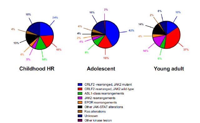
Single Tags: 

Pathology of Non-Alcoholic Fatty Liver Disease

"Non-alcoholic fatty liver disease1" by Nephron - Own work. Licensed under CC BY-SA 3.0 via Wikimedia Commons - http://commons.wikimedia.org/wiki/File:Non-alcoholic_fatty_liver_disease1.jpg#/media/File:Non-alcoholic_fatty_liver_disease1.jpg

Volume 10, Issue 1, January 2015

Dr. Anthony W.H. Chan

Associate Professor Department of Anatomical and Cellular Pathology, Prince of Wales Hospital, The Chinese University of Hong Kong

Introduction

Non-alcoholic fatty liver disease (NAFLD) is a serious global health problem and associated with over-nutrition and its related metabolic risk factors including central obesity, glucose intolerance, dyslipidaemia and hypertension. It is the most common metabolic liver disease worldwide and its prevalence in most Asian countries is similar to that in the States, Europe and Australia. About 10-45% of Asian population have NAFLD. With “westernized” sedentary lifestyle, the prevalence of NAFLD in general urban population in the mainland China is about 15%. NAFLD is even more prevalent in Hong Kong. Our recent study demonstrated that NAFLD is found in 27.3% of Hong Kong Chinese adults by using proton-magnetic resonance spectroscopy. We further realized that 13.5% of Hong Kong Chinese adults newly develop NAFLD in 3-5 years. Both prevalence and incidence of NAFLD in Hong Kong are alarmingly high. Accurate diagnosis of NAFLD is crucial to allow prompt management of patients to reduce morbidity and mortality. NAFLD is composed of a full spectrum of conditions from steatosis to steatohepatitis (NASH) and cirrhosis. Various non-invasive tests, based on clinical, laboratory and radiological tests, have been developed to assess the degree of steatosis and fibrosis in NAFLD. However, liver biopsy remains the gold standard for characterizing liver histology in patients with NAFLD, and is recommended in patients with NAFLD at high-risk of steatohepati tis and advanced fibrosis (bridging fibrosis and cirrhosis), and concurrent chronic liver disease of other aetiology. This article reviews pathological features of NAFLD and highlights some practical points for our daily diagnostic work.

Download Topic Update for Pathology of Non-Alcoholic Fatty Liver Disease

Single Tags: 

Molecular Classification and Genetic Alterations of Diffuse Large B-cell Lymphoma

"Glass ochem" by Purpy Pupple - Own work. Licensed under CC BY-SA 3.0 via Wikimedia Commons - http://commons.wikimedia.org/wiki/File:Glass_ochem.png#/media/File:Glass_ochem.png

Volume 9, Issue 2, July 2014

Dr CHOI Wai Lap

Department of Clinical Pathology Tuen Mun Hospital

Introduction

Diffuse large B-cell lymphoma (DLBCL) is the commonest subtype of non-Hodgkin lymphoma, accounting for about 30% to 40% of newly diagnosed non-Hodgkin lymphoma worldwide and in Hong Kong. DLBCL is heterogeneous in clinical presentation, morphology, immunophenotype, cytogenetics and prognos is. In the WHO Classification of Tumours of the Haematopoietic and Lymphoid Tissues published in 2008, several specific clinicopathological entities of DLBCL have been recognized, while leaving the rest to DLBCL, not otherwise specified, which is by far the most prevalent entity among the large B-cell lymphomas. In the following discussion, the term DLBCL will be used interchangeably with DLBCL, not otherwise specified.

Gene expression profiling and molecular classification of DLBCL

Gene expression profiling (GEP) is the simultaneous measurement of the transcription levels of thousands of genes to their corresponding messenger RNAs (mRNAs). GEP can be achieved by various technologies including DNA microarray, serial analysis of gene expression (SAGE) and most recently next generation sequencing (RNA-Seq). Using DNA microarray technology on DLBCL, two distinct molecular subgroups were discovered based on the similarity of their gene expression pattern with a possible cell of origin (COO): the germinal centre B-cell-like (GCB-cell-like, or abbreviated as GCB) and the activated B-cell-like(ABC-like, or abbreviated as ABC). These molecular subgroups showed significantly different survival rates when treated with conventional cyclophosphamide, doxorubicin, vincristine and prednisolone (CHOP) chemotherapeutic regimen.

Download Topic Update for Molecular Classification and Genetic Alterations of Diffuse Large B-cell Lymphoma

Single Tags: