Topic Update

Screening of DPD Deficiency before Fluoropyrimidine Chemotherapy

Screening of DPD Deficiency before Fluoropyrimidine Chemotherapy

Dr Felix Wong

Consultant Chemical Pathologist, Queen Mary Hospital

Volume 19, Issue 1, Jan 2024  (download full article in pdf)

Editorial note:

Screening of dihydropyrimidine dehydrogenase (DPD) deficiency before systemic fluoropyrimidine chemotherapy can improve safety and prevent the occurrence of the associated toxicity. Both genotyping and phenotyping approaches have been advocated. In this review, Dr Felix Wong compares and contrasts both approaches and explains the upcoming situation in Hong Kong in the near future. We welcome any feedback or suggestions. Please direct them to Dr Esther Hung of Education Committee, the Hong Kong College of Pathologists. Opinions expressed are those of the authors or named individuals, and are not necessarily those of the Hong Kong College of Pathologists.

Single Tags: 

Melioidosis: an urban outbreak in Hong Kong

Melioidosis: an urban outbreak in Hong Kong

Dr. Kristine LUK, Dr. May LEE and Dr. Wing Kin TO

Consultant Microbiologists, Department of Pathology, Princess Margaret Hospital, Hospital Authority

Volume 18, Issue 2, July 2023  (download full article in pdf)

Editorial note:

There was a significant upsurge of cases of melioidosis in Hong Kong in 2022, especially in the Kowloon region, raising public awareness to the condition. In this issue of the Topical Update, Drs. Kristine Luk, May Lee and WK To share their experience in investigating and managing the cases. We welcome any feedback or suggestion. Please direct them to Dr. Janice Lo (e-mail: janicelo@dh.gov.hk), Education Committee, The Hong Kong College of Pathologists. Opinions expressed are those of the authors or named individuals, and are not necessarily those of the Hong Kong College of Pathologists.

Single Tags: 

A Review on Complement Diagnostics

A Review on Complement Diagnostics


Volume 18, Issue 1, Jan 2023  (download full article in pdf)


Editorial note:


The complement system though commonly regarded as component of the innate immune system that protect our bodies from infection, it has increasingly evident that it has important roles in other immune surveillance and housekeeping functions, that it is involved in a wide and diverse range of clinical conditions. In this review, Dr Elaine Au provided an overview of the complement diagnostics and its clinical applications. We welcome any feedback or suggestions. Please direct them to Dr Elaine Au of Education Committee, the Hong Kong College of Pathologists. Opinions expressed are those of the authors or named individuals, and are not necessarily those of the Hong Kong College of Pathologists.


Dr Au Yuen Ling Elaine


Consultant, Division of Clinical Immunology, Department of Pathology, Queen Mary Hospital

The complement system not only as part of the innate immune system that contributes to the elimination of pathogens, and promote inflammation, it also modulates the adaptive immune response. Though its primary role is in host defense, it also serves an important role in clearance of apoptotic cells and immune complexes. Low or dysregulated activity in complement system has been described in a range of disease and pathological conditions.

The Complement system

The complement system comprises approximately 50 proteins, that are found in fluid phase or bound to cell surface (2). The central complement reaction involves the cleavage of C3 into C3b and C3a, which is promoted by the C3 convertase. Collectively, there are three activation pathways forming the C3 convertase. The classical pathway (CP) is triggered by the immune complexes, while the lectin pathway (LP) is triggered by the binding of mannan-binding lectin (MBL) or ficolins to carbohydrates or pathogen-associated molecular patterns. Both activation of CP and LP would lead to the formation of C4b2a as C3 convertase. On the other hand, in the alternative pathway (AP), there is a constant low-grade hydrolysis of C3, that binds factor B and cleaves factor D to generate a fluid phase C3 convertase, that is self- limited in healthy state. However, the AP will be activated and amplified through binding of the cleaved C3 to pathogens or altered tissues. Hence, AP helps to amplify complement activation initiated from CP and LP. The pathways converge in a common pathway to form the membrane attack complex (C5b-9). In addition, the cleavage of C3 and C5 generates C3a and C5 a, that act as anaphylatoxins, while the target bound C3 fragments (C3b, iC3b, C3d, g) facilitate phagocytosis.

The complement activation is delicately controlled by multiple soluble and membrane bound regulators. Factor H, C4b binding protein, the membrane proteins complement receptor 1 CR1 (CD35), decay acceleration factor (CD55), and membrane cofactor protein MCP (CD46), act as cofactors for plasma proteinase factor I, accelerating the decay of convertases. In addition, CD59 and C1 inhibitor regulate the C5b-9 complex and the C1 complex respectively.


Examples of complement diagnostics indications and associated disease conditions

A broad spectrum of clinical conditions is associated with complement deficiencies or its overactivation / dysregulation. The clinical consequences can be broadly categorized into three areas. 1) susceptibility to infection, 2) autoimmunity and 3) defects in controlling and limiting complement activation.


Infection susceptibility

In general, complement deficiencies are associated with increased risk of infections, especially encapsulated bacterial infections, most commonly Pneumococci, Hemophilus etc. In particular, individuals suffering from deficiencies in the terminal components (C5-C9) or properdin are susceptible to Neisseria infections. Hence, complement studies are indicated in the workup of young individuals suffering from recurrent infections (e.g. recurrent sinopulmonary infections, meningitis, etc), especially in recurrent infections caused by encapsulated bacteria. Nevertheless, primary component deficiency is rare, and most of these conditions are autosomal recessive (X-linked inheritance in properdin deficiency) (1).


Autoimmune diseases

Deficiency in early components of the CP, is frequently associated with lupus like autoimmune conditions. The associations range from 10% prevalence of lupus like conditions in C2 deficiency, to C1r/s (57% prevalence), C4 (75% prevalence) and C1q (90% prevalence) (2). These deficiencies can be confirmed in genetic studies and components measurement. Overall, primary deficiency is relatively uncommon. More often, lupus and other autoimmune immune complex diseases causes secondary complement components deficiency as consumption due to the immune complex activation. The component levels, e.g. C3 and C4 levels, are commonly employed in the workup and disease activity monitoring in these conditions. In some occasions, measuring autoantibodies, such as anti-C1q antibody in hypocomplementemic urticarial vasculitis syndrome (HUVS) and lupus, is useful for diagnosis and prognostication.


C3 nephropathy and Thrombotic microangiopathy (TMA)

Uncontrolled AP activation may result in a number of kidney diseases and systemic conditions. C3 glomerulopathy comprises C3 glumoerulonephritis (C3GN) and dense-deposit disease (DDD), is a pathological condition defined by predominant C3 accumulation, with absent or scantly immunoglobulin deposition. Atypical post infectious glomerulonephritis also falls in the continuum of C3 GN and DDD (3). In these conditions, underlying predisposition, be it genetic or acquired, may not be clinically evident until a triggering event, such as infection or pregnancy, that unfold the complement dysregulation. Besides genetic predisposition, presence of autoantibodies, e.g. C3 nephritic factor (C3 Nef), anti-factor H, have been observed in some patients. C3Nef are autoantibodies that bind to components of AP convertase, prolonging its functional half-life, leading to continuous C3 activation and consumption, with lowish CP and AP studies. Factor H has important role in the regulation of complement activation. In some patients, they are predisposed to the disease due to Factor H dysfunction caued by mutation or anti-Factor H. Useful workup for C3 nephropathy includes the complement pathways, components and activation products studies, testing for plasma cells disorders, determination of autoantibodies (C3 Nef, anti-factor H), along with gene panel (C3, CFH, CFI, CFB, CFHR1-5) testing (3).

aHUS is a primary TMA, that is characterized by uncontrolled AP activation, presenting with microangiopathic hemolytic anaemia, thrombocytopenia and acute renal failure. The dysregulated AP could be caused by mutations of complement regulators, most commonly factor H, and in around 6-10% of cases, by the presence of anti- factor H (4). Initial workup includes investigations to exclude other co-existing medical conditions associated with HUS or other forms of TMA. Similar to the workup of C3GN, checking the complement pathways, components and activation products, along with anti-factor H and genetic testing (C3, CFH, CGI, CFB, MCP, CFHR1-5, THBD, DGKE) are useful.

TMA leads to generalized endothelial dysfunction, that can progress to multiorgan injury. Apart from primary causes, some disease or medical conditions may predispose to TMA. In particular transplant associated TMA (TA-TMA) has been an important clinical entity, that carries high mortality and morbidity. Recent literature has shown that complement pathway dysregulation may play a role in the process. The pathogenesis in TA-TMA is complex, that multifactorial factors contribute to the endothelial injury and pathological process. Complications related to transplant, including GVHD or infections, may also stimulate the complement pathways. Complement blockage therapy, e.g. eculizumab, is useful in managing complex cases. After workup to exclude other potential differential diagnoses, risk assessment is important. Although not all patients with TA-TMA will have elevated sC5b-9, patients with elevation are at increased risk of death from TA-TMA (5). Hence, the activation product measurement has been used as risk stratification for consideration of complement blockade therapy (4,6).


Paroxysmal Noctural Hemoglobuinuria (PNH)

PNH is a rare acquired disorder, that patients suffered from hemolysis with acute exacerbations, leading to anaemia, bone marrow failure and increased risk of thrombosis. PNH arises from an expanded clonal proliferation of hematopoietic cells with somatic mutations of the X chromosomal gene PIG-A. Lack of PIG-A resulted in inability to bind GPI-anchored proteins, including the membrane bound complement regulators, DAF and CD59. As a result, cells having the mutation are susceptible to complement mediated intravascular haemolysis. Assessing the surface expression of CD55 and CD59 is helpful for the diagnosis.


Inherited and Acquired C1 inhibitor deficiency

Hereditary angioedema (HAE) and acquired angioedema (AAE), are rare diseases caused by C1 inhibitor deficiency. As a result, unregulated bradykinin formation leads to angioedema. HAE is an autosomal dominant condition, with majority of cases suffered from reduced concentration (Type I) or less commonly, reduced function (Type II), of C1 inhibitor. Some patients may have similar clinical presentations as HAE cases, but as an acquired condition due to the presence of autoantibodies against C1 inhibitor. These patients usually presented at an older age, and may have underlying hematological malignancies or autoimmune conditions as predisposition. The diagnosis of HAE is based on C1 inhibitor and C4 measurement. It is important to include both antigenic and functional assays for C1 inhibitor, since around 15% of cases may have normal or elevated dysfunctional C1 inhibitor protein (Type II). Furthermore, serum C1q concentrations can be used to differentiate HAE from acquired angioedema (AAE) as the latter is characterized by decreased C1q antigen concentration and autoantibodies against C1-INH. Genetic analysis for SERPING1 variants status may also help in the workup.


Monitoring of Complement Regulatory Drugs

In recent years, drugs targeting complement activation has been in clinical use. Eculizumab is the first approved complement inhibitor, that it is a humanized monoclonal antibody that hinder C5 proteolytic activation, inhibit the generation of C5a and the initiation of the membrane attack complex C5b-9, through its binding to the C5. Eculizumab is approved in the treatment of PNH, aHUS and refractory myasthenia gravis. Complement studies, such as CH50/ AH 50, and activation products (sC5b-9), have been employed in the treatment monitoring (7). In some specialized laboratory, C5 function may also be tested. The best time to monitor the therapy is at trough, immediately before the next dose. With effective drug treatment, CH50/AH50 and C5 function will be low. The activation products will also be suppressed.


Complement assays

The assays used in complement assessment can be broadly divided into 1) screening assays of total functional complement activity, 2) quantification of individual components, 3) quantitation of activation products 4) detection of autoantibodies against the complement components 5) assessing cell surface expression or tissue deposition of complement proteins/ breakdown products, 6) genetic assays.

Apart from the rare primary component deficiency, complement is associated in a number of disease conditions (such as infections, sepsis, malignancy, immune complex diseases, etc) by activation via different pathways. When a component is activated in vivo, the component is taken up by receptors on leukocytes or Kupffer cells. This results in secondary deficiency as consumption. Note that in complement studies, some assays are sensitive to in-vitro activation. Consumption can also be an artifact from heat labile nature of the complement proteins combined with delayed freezing of specimen after sample collection. Overall, the specificity of single complement test is low. Assessing several markers of the pathways and careful interpretation of results as a whole, is useful. In some situations, complementary use of genetic tests may help in cases suspecting primary in nature.

Since EDTA is able to inhibit complement activation in vitro, it is commonly used for quantification of complement components, in particular for activation products. Since heparin and citrate are insufficient inhibitors of complement activation, these are not suitable. Serum, on the other hand, is used for complement function and autoantibodies assessment. Plasma and serum received for complement assays should be separated within 2 hours from collection and frozen at -70 degree Celsius (4). Careful attention to the pre analytical steps and storage is crucial in complement studies.


Screening assays for total functional complement activity

The main indication for total complement function screen is to detect complement deficiencies. Such deficiencies can be genetic (primary), acquired (secondary, e.g. to consumption after pathway activation), or as a consequence of treatment. These tests reflect the total amount of active complement component present in a freshly sampled serum, and reflect the potential of the serum sample to achieve full activation in vitro after addition of activator. The traditional assays used are CH50 and AH50, based on studying the lysis of antibody sensitized sheep erythrocytes (CH50 for the CP activity) and the lysis of untreated rabbit erythrocytes (AH50 for the AP activity). The lysis of erythrocytes correlates with the formation of the terminal membrane attack complex downstream of the pathways’ activation. The results are usually expressed as reciprocal dilutions of the sample required to produce 50% lysis. Besides the traditional assays, a variety of modified methods based on the hemolytic assay were done in different centers. The functional screen can also be tested by measuring the deposition of activation products (ELISA detecting C9 neoepitope generated in terminal complex formation) upon activation of the serum with immobilized complement activating substances on a microtiter plate. Targeted molecules for each pathway are coated in wells of the microtiter plates; Ig M for CP, mannan /acetylated bovine serum albumin for LP and LPS for AP. (8)

In general, the pathway screens may provide some hint to the underlying disease process. Absent/low AH50 with normal CH50 suggests alternative pathway component deficiency, while absent/low CH50 with normal AH 50 suggests early classical pathway components (C1, C2, C4) deficiency. Absent/low results in both AH50 and CH50 suggests a deficiency affecting common components (C3, C5, C6, C7, C8, C9) shared in both pathways or complement consumption. Further investigations, including quantitation of individual components, would be helpful. In the settings of multiple components deficiency, consumptive depletion is likely.


Quantitation of individual components

In cases where the screening assays indicating a complement deficiency, quantitation of individual components and interpreting the results as a profile is useful to further delineate the affected pathways and pathogenesis.

Measurement of complement components is commonly done by immunoprecipitation assays with polyclonal antibodies against the protein of choice, e.g. nephelometry and turbidimetry. Other assays, such as gel precipitation assays or enzyme immunoassays were also used. Overall, these assays are relatively robust, however, do not provide information on the conformation or activation status in vivo.


Quantitation of activation products

Abnormal total complement functional screen could be due to primary deficiency or deficiency secondary to consumptive loss. Measurement of individual components level is not able to distinguish between primary from secondary loss. On the other hand, in vivo complement activation in acute phase reaction may not always lead to low components measurement despite ongoing consumption. Hence, quantitation of activation products would be helpful in the assessment of complement activation. Among the activation products available for measurement, detection of the soluble form of the terminal complement complex (sC5b-9), is the most promising screen for complement activation. The terminal complex reflects the activation to the final stage of the three pathways. Moreover, sC5b-9 has a relatively long in vivo half-life (60 mins), compared to other activation products, and is more stable with respect to in vitro activation compared to early components fragments (1,4). Overall, these activation markers can be rapidly produced by complement activation in vitro, therefore, proper sample collection and handling is important.


Autoantibodies against complement components

Autoantibodies to complement components have been linked to a number of disease conditions. The pathogenesis is often caused by the dysregulation of complement activation, as in the case of C3NeF and anti-Factor H. Occasionally, it may be affecting non-complement pathway, as in the case of anti-C1 inhibitor related angioedema, that it is due to inefficient inhibition of the kallikrein-kinin system and bradykinin release (4).

Most often, these autoantibodies could be detected by enzyme immunoassays. Functional assays were also helpful in the assessment. For example, in C3 Nef detection, a hemolytic assay that utilizes unsensitized sheep erythrocytes, or assay detecting fluid-phase C3 conversion after incubation of patient serum with normal serum at 37degree Celsius, were commonly used for the C3 Nef activity detection (9).

Assessing cell surface expression or tissue deposition of complement proteins/ breakdown products Measuring complement components and activation products directly on cell surface provides valuable information for the workup. For example, examining the deposition of various complement components in the glomeruli and peritubular capillary is useful for glomerulopathies assessment. Furthermore, studying the expression of membrane bound regulators is also helpful in some conditions, such as the use of flow cytometry assessment of CD55 and CD59 on blood cells in the diagnosis of PNH.


Genetic assays

With the advances in molecular diagnostics, complementary use of molecular diagnostics with traditional assays, has been increasingly employed in cases suspecting primary deficiency of complement factors or regulators. For example, gene panels study has been recommended in the workup of aHUS and C3 glomerulonephritis (3, 10-11).


Conclusion

With the vast and constantly growing knowledge in various disease process, along with expanding indications and emerging treatment options in complement mediated disorders, the application of complement diagnostics has been broadened and not limited to diagnosing rare primary genetic entities only. However, many of these assays remains highly subspecialized with limited availability, lack of standardization and complex interpretations. Careful standardization and close international collaborations and experience sharing, would be important for both the laboratory development and clinical applications in the field.


References

  1. Kirschfink M, Mollnes TE. Modern complement analysis. Clin Diagn Lab Immunol. 2003 Nov;10(6):982-9.
  2. Pickering MC, Botto M, Taylor PR, Lachmann PJ, Walport MJ. Systemic lupus erythematosus, complement deficiency, and apoptosis. Adv Immunol. 2000;76:227-324.
  3. Angioi A, Fervenza FC, Sethi S, Zhang Y, Smith RJ, Murray D, Van Praet J, Pani A, De Vriese AS. Diagnosis of complement alternative pathway disorders. Kidney Int. 2016 Feb;89(2):278-88.
  4. Ekdahl KN, Persson B, Mohlin C, Sandholm K, Skattum L, Nilsson B. Interpretation of Serological Complement Biomarkers in Disease. Front Immunol. 2018 Oct 24;9:2237.
  5. Jodele S, Davies SM, Lane A, Khoury J, Dandoy C, Goebel J, Myers K, Grimley M, Bleesing J, El-Bietar J, Wallace G, Chima RS, Paff Z, Laskin BL. Diagnostic and risk criteria for HSCT-associated thrombotic microangiopathy: a study in children and young adults. Blood. 2014 Jul 24;124(4):645-53.
  6. Jodele S, Dandoy CE, Lane A, Laskin BL, Teusink-Cross A, Myers KC, Wallace G, Nelson A, Bleesing J, Chima RS, Hirsch R, Ryan TD, Benoit S, Mizuno K, Warren M, Davies SM. Complement blockade for TA-TMA: lessons learned from a large pediatric cohort treated with eculizumab. Blood. 2020 Mar 26;135(13):1049-1057.
  7. Ricklin D, Barratt-Due A, Mollnes TE. Complement in clinical medicine: Clinical trials, case reports and therapy monitoring. Mol Immunol. 2017 Sep;89:10-21.
  8. Mollnes TE, Lea T, Frøland SS, Harboe M. Quantification of the terminal complement complex in human plasma by an enzyme-linked immunosorbent assay based on monoclonal antibodies against a neoantigen of the complex. Scand J Immunol. 1985 Aug;22(2):197-202.
  9. Nilsson B, Ekdahl KN. Complement diagnostics: concepts, indications, and practical guidelines. Clin Dev Immunol. 2012;2012:962702.
  10. Goodship TH, Cook HT, Fakhouri F, Fervenza FC, Frémeaux-Bacchi V, Kavanagh D, Nester CM, Noris M, Pickering MC, Rodríguez de Córdoba S, Roumenina LT, Sethi S, Smith RJ; Conference Participants. Atypical hemolytic uremic syndrome and C3 glomerulopathy: conclusions from a "Kidney Disease: Improving Global Outcomes" (KDIGO) Controversies Conference. Kidney Int. 2017 Mar;91(3):539-551.
  11. Loirat C, Fakhouri F, Ariceta G, Besbas N, Bitzan M, Bjerre A, Coppo R, Emma F, Johnson S, Karpman D, Landau D, Langman CB, Lapeyraque AL, Licht C, Nester C, Pecoraro C, Riedl M, van de Kar NC, Van de Walle J, Vivarelli M, Frémeaux-Bacchi V; HUS International. An international consensus approach to the management of atypical hemolytic uremic syndrome in children. Pediatr Nephrol. 2016 Jan;31(1):15-39.
Single Tags: 

Molecular diagnostics for breast cancer

Molecular diagnostics for breast cancer


Volume 17, Issue 2, July 2022  (download full article in pdf)


Editorial note:


New molecular techniques have contributed to the ever-expanding armamentarium for breast cancer diagnosis, treatment and prognostication. Since the molecular classification of breast cancer was established, pathologists have been using immunohistochemistry and DNA sequencing techniques to routinely grade and subtype breast cancer. RNA expression profiling using various platforms such as microarrays, quantitative PCR and Nanostring has also been used to guide patient treatment in early diseases. This topical update provides a concise review on the current diagnostic and prognostic modalities in breast cancer management. We welcome any feedback or suggestions. Please direct them to Dr. Alvin Cheung of Education Committee, the Hong Kong College of Pathologists. Opinions expressed are those of the authors or named individuals, and are not necessarily those of the Hong Kong College of Pathologists.


Dr. Alvin Ho-Kwan Cheung1 and Dr. Karen Ka-Wan Yuen2

  1. Department of Anatomical and Cellular Pathology, Prince of Wales Hospital, the Chinese University of Hong Kong
  2.  
  3. Department of Clinical Pathology, North District Hospital


Introduction

Since the seminal report on breast cancer classification in 2000[1], increased understanding in the molecular biology of breast cancer has led to numerous immunohistochemical markers and molecular panels used as adjunct biomarkers. These biomarkers mainly serve the following purposes: As prognostic markers, to gauge the likelihood of a clinical event, disease recurrence or progression; as predictive markers, to assess the likelihood of favourable or unfavourable effect from exposure to a medical product or a therapeutic agent[2]. In this review, the classical biomarkers of breast cancer will be briefly discussed, followed by a more detailed elaboration of molecular panels which are based on DNA alterations and gene expression levels. 


Hormonal receptor and proliferative index markers

Unlike other cancers, the molecular classification of breast cancer (luminal A/B, HER2 positive, and basal-like cancers) have been translated well to the clinic[3], and immunohistochemical markers have been established to facilitate such classification without resorting to molecular methods[4, 5]. Some authorities believe that normal-like breast cancer are an artifact of contamination by normal cells[6, 7]. The Estrogen Receptor (ER) and Progesterone Receptor (PR) are predictive biomarkers for endocrine therapy[8]. ER or PR-expressing tumours tend to have a better outcome than those lacking the receptors.

The expression of Human Epidermal growth factor Receptor 2 (HER2) defines the molecular basis of the “HER2-positive” group of cancer. They account for slightly less than 20% of breast cancers, and have a worse prognosis compared to ER+/PR+/HER2- cancers[9]. It serves as a therapeutic target for trastuzumab and pertuzumab. While ER and PR are routinely detected by immunohistochemistry (IHC), HER2 expression can be detected by IHC, Dual in situ hybridization (DISH) or Fluorescence in situ hybridization (FISH)[10].


The proliferation marker Ki-67 serves as a useful adjunct investigation in the grading of breast cancer[11]. Calculated as the percentage of nuclear staining in cancer cells, the prognosis is said to be better when Ki-67 is 30% for early disease[12].


Molecular tests at the DNA level

Some genetic aberrations in breast cancer are worth mentioning because they may be susceptible to targeted therapy and can predict treatment response. PIK3CA mutation occurs in about 36% of breast cancer[13, 14]. In advanced or metastatic hormonal receptor-positive cancer, or in patients with disease progression on endocrine-based regimen, combination therapy with the PI3K inhibitor, alpelisib, together with fulvestrant may be a treatment option if there is PIK3CA mutation[15]. The mutation can be detected by the companion diagnostic kit Therascreen, with Sanger sequencing, or with next generation sequencing. In secretory carcinoma, NTRK fusion is targetable by larotrectinib or entrectinib[16]. The presence of translocation can be detected with immunohistochemistry, next generation sequencing (NGS), Reverse-transcriptase (RT)-PCR or FISH. In non-secretory type breast cancers, NTRK fusion is very rare[17], such that the routine testing of this gene is unnecessary. For triple-negative breast cancer, BRCA aberrations can be present in about 6.5-34% of the cases[18]. The BRCA proteins constitute a part of the homologous recombination repair pathway. They are encoded by relatively large genes, with BRCA1 being present on chromosome17q21, having 23 exons; and BRCA2 on chromosome 13q13.1, having 27 exons. The incidence of aberrations is markedly higher among Ashkenzi Jews (2.5%) than the general population (0.1%)[19]. BRCA-mutated tumour highly depends on PARP, another DNA repair protein, to maintain the tumour genome integrity. Therefore, PARP inhibitor therapy are useful in BRCA-mutated tumours, and this treatment approach is termed a “synthetic lethality”[20].  Due to the size of these genes, NGS would be the preferred detection platform, while multiplex ligation-dependent probe amplification (MLPA) is also suitable[21]. 


For other advanced cancer or triple negative breast cancer, immune checkpoint inhibitor may be indicated in some patients. Besides testing for PD-L1 expression by the companion diagnostic kits for atezolizumab and pembrolizumab, some data support the testing for microsatellite instability (MSI) and tumour mutation burden (TMB) as well[22]. MSI-high breast cancer may be treated with pembrolizumab, as are tumours with high TMB as assessed by the FoundationOne companion diagnostic or other NGS platforms[23].


Molecular tests at the RNA expression level

Oncotype Dx

Oncotype Dx was launched in year 2004. It involves mRNA extraction from formalin-fixed paraffin-embedded (FFPE) tissues[24]. The detection panel includes 21 genes (16 cancer-related genes and 5 reference genes), and the detection platform is by quantitative-PCR (qPCR). The test had been studied in several trials, including the NASBP trial (National Surgical Adjuvant Breast and Bowel Project)[25], TAILORx trial (including 10273 women), and TxPONDER trial (5018 women)[26]. The test generates a recurrence score (RS) in the range of 0-100. In the TAILORx trial, patients of age >50 years had a substantial benefit from chemotherapy when RS >=26, whereas younger patients may be benefited when RS >=16.

MammaPrint

MammaPrint was launched in 2007. It consists of a 70-gene microarray, which accepts both fresh frozen or FFPE tissue for testing. It categorizes patients into “High risk” or “Low risk”. The MINDACT trial included 6693 patients and the RASTER trial included 427 patients for this test[27, 28]. There are some preliminary data to suggest systemic treatment can be recommended for the patients in the “High risk” group.

Blueprint

The Blueprint assay was developed by the same company as Mammaprint, and the test can be used together with MammaPrint. It involves a 80-gene panel, and serves to categorize tumour into luminal-A, luminal-B, HER2, or basal subtypes. Although this may overlap with the objective of IHC study described above, one important difference is that the luminal A and B groups can be associated with a different chemosensitivity and prognosis according to the Blueprint schema. Particularly, in the luminal B, Her2, and basal subtypes, chemotherapy can be beneficial to some patients with an improved survival. In contrast, for the luminal A group, the benefit for chemotherapy is not pronounced[29].

Prosigna (PAM50)

The Prosigna assay was launched in 2013. Following RNA extraction from FFPE tissue, the expression of a panel of 50 genes are detected by the NanoString “nCounter” platform[30]. This test is indicated for post-menopausal patients. Two large trials were conducted, including The ABCSG-8 study (Austrian Breast and Colorectal Cancer Study Group 8) and TransATAC study (translational arm of the anastrozole or tamoxifen alone or combined)[31]. While a scoring scheme of 0-100 is used, the risk stratification is different depending on the lymph node status. For node-negative cancers, they are classified as low (0-40), intermediate (41-60), or high (61-100) risk; as for node-positive cancers, they are classified as low (0-40) or high (41-100) risk. The suggested treatment for low risk disease is hormonal therapy alone, while for high risk disease, chemotherapy in addition to hormonal therapy may be beneficial.

The Breast Cancer Index

The Breast Cancer Index was launched in 2008[32]. As an RT-PCR assay on FFPE tissue, it features a 11-gene panel with two major testing endpoints: Whether there is a benefit of extended endocrine therapy (for 5 years), and the risk of recurrence 5 to 10 years after diagnosis. The ratio of expression between estrogen signaling pathway genes HOXB13 and IL17BR (H/I ratio) is an important parameter, as in the MA.17 trial, high H/I indicated a higher risk of late recurrence and a benefit from extended letrozole therapy. Another trial, the aTTom study, included H/I high patients for an extended therapy and found up to 15% reduction in recurrence risk[33, 34]. The test results for the Breast Cancer Index are simple enough to be interpreted even by patients, with “Yes” and “No” to the question of whether extended endocrine therapy is beneficial, and recurrence risk in percent to report the chance of late distant recurrence.

Comparisons between test modalities

When the included genes are compared, it is noted that the Oncotype Dx and PAM50 panels have the most overlap. 11 genes are in common for Oncotype Dx and PAM50, for example BCL2, CCNB1, MMP11, which are markers for apoptosis, cell cycle, and tumour invasiveness[35]. Interestingly, for the 70 genes included in Mammaprint, only one gene, SCUBE2, overlaps with Oncotype DX, and two genes, MELK and ORC6L, overlap with Prosigna PAM50[35]. It remains to be studied whether the results in one test can be correlated with another test, but some key differences are still worth to be noted. For hormonal receptor positive stage I-II invasive breast cancer, all the tests have some use for prognostication. However, concerning whether chemotherapy is recommended, only Oncotype DX has an established predictive value, while there is insufficient evidence for Mammaprint, Blueprint and Prosigna [36]. The Breast Cancer Index has predictive value for extended endocrine therapy. Some efforts have also been taken to translate some of these tests to hormonal receptor positive DCIS. Oncotype DX DCIS and DCISionRT have some use in patient prognostication, however, both tests have insufficient evidence to guide chemotherapy[37, 38].

For the regulatory status, Oncotype DX has been included in the NCCN/ASCO guidelines for the management of breast cancer patients. As for Mammaprint and Prosigna, these kits have been FDA-cleared for specific clinical settings. Logistically, both Oncotype DX and Mammaprint require end users to deliver specimens to a central laboratory for testing. Prosigna is available in a kit format for local laboratories to perform the test.


Conclusions and future perspectives

Unlike most other cancer types, RNA expression profiling has found remarkable translational use in breast cancer treatment. This can be attributed to an increased understanding of molecular classifications, hormonal receptor functions and breast cancer biology. While panels including other RNA expression signatures can be expected to emerge, it is important to understand the indications and differences for each testing system, as an increased number of testing options can be confusing to patients, while contradicting results among platforms can complicate the interpretation. Because RNA expression level has an inherent variability among patients, the subgrouping of patients into risk groups may not be ideal, and some patients may be placed in the wrong group using only one particular panel. Hopefully, with further elucidation of the breast cancer genome, novel molecular targets based on DNA alterations can be uncovered, as the presence of a particular mutation or translocation is a more consistent marker of susceptibility to targeted therapy. As we enter the era of personalized medicine, histologic assessments, immunohistochemical studies such as hormonal receptors and PD-L1 status, and molecular diagnostics can be expected to go hand in hand in the formulation of management plans and prognostication in breast cancer patients.


Reference

  1. Perou CM, Sørlie T, Eisen MB, Van De Rijn M, Jeffrey SS, Rees CA, et al. Molecular portraits of human breast tumours. nature. 2000;406(6797):747-52.
  2. Califf RM. Biomarker definitions and their applications. Experimental Biology and Medicine. 2018;243(3):213-21.
  3. Sotiriou C, Pusztai L. Gene-expression signatures in breast cancer. New England Journal of Medicine. 2009;360(8):790-800.
  4. Goldhirsch A, Winer EP, Coates A, Gelber R, Piccart-Gebhart M, Thürlimann B, et al. Personalizing the treatment of women with early breast cancer: highlights of the St Gallen International Expert Consensus on the Primary Therapy of Early Breast Cancer 2013. Annals of oncology. 2013;24(9):2206-23.
  5. Cheang MC, Martin M, Nielsen TO, Prat A, Voduc D, Rodriguez‐Lescure A, et al. Defining breast cancer intrinsic subtypes by quantitative receptor expression. The oncologist. 2015;20(5):474-82.
  6. Tsang J, Tse GM. Molecular classification of breast cancer. Advances in anatomic pathology. 2020;27(1):27-35.
  7. Peppercorn J, Perou CM, Carey LA. Molecular subtypes in breast cancer evaluation and management: divide and conquer. Breast Cancer. 2007:125-42.
  8. Prat A, Baselga J. The role of hormonal therapy in the management of hormonal-receptor-positive breast cancer with co-expression of HER2. Nature Clinical Practice Oncology. 2008;5(9):531-42.
  9. Cooke T, Reeves J, Lanigan A, Stanton P. HER2 as a prognostic and predictive marker for breast cancer. Annals of oncology. 2001;12:S23-S8.
  10. Kurozumi S, Padilla M, Kurosumi M, Matsumoto H, Inoue K, Horiguchi J, et al. HER2 intratumoral heterogeneity analyses by concurrent HER2 gene and protein assessment for the prognosis of HER2 negative invasive breast cancer patients. Breast cancer research and treatment. 2016;158(1):99-111.
  11. Cheang MC, Chia SK, Voduc D, Gao D, Leung S, Snider J, et al. Ki67 index, HER2 status, and prognosis of patients with luminal B breast cancer. JNCI: Journal of the National Cancer Institute. 2009;101(10):736-50.
  12. Nielsen TO, Leung SCY, Rimm DL, Dodson A, Acs B, Badve S, et al. Assessment of Ki67 in breast cancer: updated recommendations from the international Ki67 in breast cancer working group. JNCI: Journal of the National Cancer Institute. 2021;113(7):808-19.
  13. Anderson EJ, Mollon LE, Dean JL, Warholak TL, Aizer A, Platt EA, et al. A systematic review of the prevalence and diagnostic workup of PIK3CA mutations in HR+/HER2–metastatic breast cancer. International Journal of Breast Cancer. 2020;2020.
  14. Deng L, Zhu X, Sun Y, Wang J, Zhong X, Li J, et al. Prevalence and prognostic role of PIK3CA/AKT1 mutations in Chinese breast cancer patients. Cancer Research and Treatment: Official Journal of Korean Cancer Association. 2019;51(1):128.
  15. André F, Ciruelos E, Juric D, Loibl S, Campone M, Mayer I, et al. Alpelisib plus fulvestrant for PIK3CA-mutated, hormone receptor-positive, human epidermal growth factor receptor-2–negative advanced breast cancer: final overall survival results from SOLAR-1. Annals of Oncology. 2021;32(2):208-17.
  16. Ricciuti B, Genova C, Crinò L, Libra M, Leonardi GC. Antitumor activity of larotrectinib in tumors harboring NTRK gene fusions: a short review on the current evidence. OncoTargets and therapy. 2019;12:3171.
  17. Remoué A, Conan‐Charlet V, Bourhis A, Flahec GL, Lambros L, Marcorelles P, et al. Non‐secretory breast carcinomas lack NTRK rearrangements and TRK protein expression. Pathology International. 2019;69(2):94-6.
  18. Hartman AR, Kaldate RR, Sailer LM, Painter L, Grier CE, Endsley RR, et al. Prevalence of BRCA mutations in an unselected population of triple‐negative breast cancer. Cancer. 2012;118(11):2787-95.
  19. Warner E, Foulkes W, Goodwin P, Meschino W, Blondal J, Paterson C, et al. Prevalence and penetrance of BRCA1 and BRCA2 gene mutations in unselected Ashkenazi Jewish women with breast cancer. Journal of the National Cancer Institute. 1999;91(14):1241-7.
  20. Cong K, Peng M, Kousholt AN, Lee WTC, Lee S, Nayak S, et al. Replication gaps are a key determinant of PARP inhibitor synthetic lethality with BRCA deficiency. Molecular cell. 2021;81(15):3128-44. e7.
  21. Wallace AJ. New challenges for BRCA testing: a view from the diagnostic laboratory. European Journal of Human Genetics. 2016;24(1):S10-S8.
  22. Rozenblit M, Huang R, Danziger N, Hegde P, Alexander B, Ramkissoon S, et al. Comparison of PD-L1 protein expression between primary tumors and metastatic lesions in triple negative breast cancers. Journal for Immunotherapy of Cancer. 2020;8(2).
  23. Sivapiragasam A, Ashok Kumar P, Sokol ES, Albacker LA, Killian JK, Ramkissoon SH, et al. Predictive biomarkers for immune checkpoint inhibitors in metastatic breast cancer. Cancer medicine. 2021;10(1):53-61.
  24. Carlson JJ, Roth JA. The impact of the Oncotype Dx breast cancer assay in clinical practice: a systematic review and meta-analysis. Breast cancer research and treatment. 2013;141(1):13-22.
  25. McVeigh TP, Hughes LM, Miller N, Sheehan M, Keane M, Sweeney KJ, et al. The impact of Oncotype DX testing on breast cancer management and chemotherapy prescribing patterns in a tertiary referral centre. European Journal of Cancer. 2014;50(16):2763-70.
  26. Lafitte E, Sabatier R. Genomic/transcriptomic signatures in breast cancer. A review of three prospective studies. Innovations & Thérapeutiques en Oncologie. 2022;8(1):5-11.
  27. Metzger O, Cardoso F, Poncet C, Desmedt C, Linn S, Wesseling J, et al. Clinical utility of MammaPrint testing in invasive lobular carcinoma: results from the MINDACT phase III trial. European Journal of Cancer. 2020;138:S5-S6.
  28. Drukker CA, Bueno‐de‐Mesquita J, Retèl VP, van Harten WH, van Tinteren H, Wesseling J, et al. A prospective evaluation of a breast cancer prognosis signature in the observational RASTER study. International journal of cancer. 2013;133(4):929-36.
  29. Mittempergher L, Delahaye LJ, Witteveen AT, Snel MH, Mee S, Chan BY, et al. Performance characteristics of the BluePrint® breast cancer diagnostic test. Translational oncology. 2020;13(4):100756.
  30. Wallden B, Storhoff J, Nielsen T, Dowidar N, Schaper C, Ferree S, et al. Development and verification of the PAM50-based Prosigna breast cancer gene signature assay. BMC medical genomics. 2015;8(1):1-14.
  31. Lænkholm A-V, Jensen M-B, Eriksen JO, Roslind A, Buckingham W, Ferree S, et al. Population-based study of Prosigna-PAM50 and outcome among postmenopausal women with estrogen receptor-positive and HER2-negative operable invasive lobular or ductal breast cancer. Clinical breast cancer. 2020;20(4):e423-e32.
  32. Ma X-J, Salunga R, Dahiya S, Wang W, Carney E, Durbecq V, et al. A five-gene molecular grade index and HOXB13: IL17BR are complementary prognostic factors in early stage breast cancer. Clinical cancer research. 2008;14(9):2601-8.
  33. Noordhoek I, Treuner K, Putter H, Zhang Y, Wong J, Kranenbarg EM-K, et al. Breast cancer index predicts extended endocrine benefit to individualize selection of patients with HR+ early-stage breast cancer for 10 years of endocrine therapy. Clinical Cancer Research. 2021;27(1):311-9.
  34. Bartlett J, Sgroi D, Treuner K, Zhang Y, Ahmed I, Piper T, et al. Breast Cancer Index and prediction of benefit from extended endocrine therapy in breast cancer patients treated in the Adjuvant Tamoxifen—To Offer More?(aTTom) trial. Annals of Oncology.
  35. 2019;30(11):1776-83.
  36. Pennock ND, Jindal S, Horton W, Sun D, Narasimhan J, Carbone L, et al. RNA-seq from archival FFPE breast cancer samples: molecular pathway fidelity and novel discovery. BMC medical genomics. 2019;12(1):1-18.
  37. Duffy M, Harbeck N, Nap M, Molina R, Nicolini A, Senkus E, et al. Clinical use of biomarkers in breast cancer: Updated guidelines from the European Group on Tumor Markers (EGTM). European journal of cancer. 2017;75:284-98.
  38. Nofech-Mozes S, Hanna W, Rakovitch E. Molecular evaluation of breast ductal carcinoma in situ with oncotype DX DCIS. The American journal of pathology. 2019;189(5):975-80.
  39. Shah C, Bremer T, Cox C, Whitworth P, Patel R, Patel A, et al. The Clinical Utility of DCISionRT® on Radiation Therapy Decision Making in Patients with Ductal Carcinoma In Situ Following Breast-Conserving Surgery. Annals of surgical oncology. 2021;28(11):5974-84.
Single Tags: 

Measurable residual disease (MRD) for Haematological Malignancy

Measurable residual disease (MRD) for Haematological Malignancy


Volume 17, Issue 1, January 2022  (download full article in pdf)


Editorial note:


Measurable residual disease (MRD) monitoring has emerged as an important indicator for risk stratification and treatment planning in patients with haematological malignancies. In the past decade, various techniques in measuring MRD have become available in Hong Kong. In this Topical Update, Dr. YIP Sze-fai provides an overview of the current techniques available for MRD monitoring. We welcome any feedback or suggestions. Please direct them to Dr. Alvin IP of Education Committee, the Hong Kong College of Pathologists. Opinions expressed are those of the authors or named individuals, and are not necessarily those of the Hong Kong College of Pathologists.


Dr. YIP Sze-fai

Consultant Haematologist, Department of Clinical Pathology, Tuen Mun Hospital


Introduction


Measurable residual disease (MRD) describes the application of assays for detection of submicroscopic level of residual disease burden which cannot be detected by morphology. Numerous studies have observed the association of MRD level and disease prognosis. It provides an objective parameter on the tumor burden, and guide stratified treatment including the application of haemopoietic stem cell transplantation (HSCT). Its ability to monitor disease and to detect molecular relapse enables preemptive therapy to prevent frank disease relapse [1]. For all these reasons, we see an increasing use of MRD in the field of haematological malignancy.


Different technologies are used for MRD measurement


1. Multiparametric flow cytometry (MFC)

MFC is commonly used for MRD detection in acute leukaemias. At diagnosis, the leukaemia-associated immunophenotype (LAIP) of the blasts can be determined by using a multitude of fluorochrome-labeled monoclonal antibodies against different cellular markers that aids identification of the leukaemic population as well as detecting the aberrant cellular marker expression. If the LAIP was not determined at diagnosis, a different-from-normal (DfN) approach can be used to detect the abnormal cells, as well as detecting any new or disappearance of known phenotypic aberrancies [1,2]. With technological advancement, more fluorochromes are available and 8 to 12-colour panels are commonly used. Flow cytometry has the advantage of a short turnaround time which can provide timely results for clinical decision making. The sensitivity of MRD detection is at the level of 10-4 to 10-5.


2. Next generation flow (NGF) for plasma cell myeloma

Novel Euroflow-based next generation flow (NGF) approach is being developed for highly sensitive and standardized MRD detection, primarily in plasma cell myeloma, using an optimized 2-tube 8-color antibody panel [3]. The NGF approach uses tools and procedures that are developed by the EuroFlow Consortium for a standardized sample preparation, antibody panel (including the type of antibody and fluorochrome), and automatic identification of plasma cells against reference databases of normal and patient BM using Infinicyt software. The sensitivity of MRD detection is close to 10-6.


3. Quantitative polymerase chain reaction (qPCR) technique

a. Detection of leukaemia-specific fusion transcript

The MRD can be measured by detecting the amount of leukaemia-specific fusion transcripts present. The classical example is BCR-ABL1 fusion in chronic myeloid leukaemia (CML). The sensitivity is higher than that of flow cytometry, reaching the level of 10-4 to 10-6. The test is relatively easy to be performed in hospital service laboratory. The MRD is represented in a ratio of normalized copy number of the fusion transcript and the control gene transcript (e.g. ABL1). For CML monitoring, an international scale (IS) ratio is developed for standardization of results among different laboratories [4]. Yet, this method is limited to cases with targetable fusion transcripts available for detection.

b. Allele-specific oligonucleotide (ASO) qPCR for immunoglobulin (IG) or T cell receptor (TCR) gene rearrangement

ASO qPCR can be employed to detect the disease-specific sequence of rearranged IG gene or TCR gene in the sample. The sensitivity of this method is 10-4 to 10-5. It is applicable to most of the cases of acute lymphoblastic leukemia (ALL) and plasma cell myeloma as long as a disease-specific rearrangement can be determined by sequencing. Patient-specific primers would need to be designed for each case. It has a disadvantage that if there is a clonal evolution, the disease-specific rearrangement can be lost and a false-negative result can be generated.


4. Digital droplet polymerase chain reaction (ddPCR)

In ddPCR, the sample is compartmentalized into very large number of separate small volume reactions. As a result, either zero or one target molecule could be detected inside any individual reaction. Thermal cycling would be performed to endpoint using same primer and probes as qPCR. Any target-containing compartments will become brightly fluorescent while compartments without targets will have only background fluorescence. Total number of ‘positive’ reactions is equal to the number of original target molecules in the entire volume, and the total number of reactions multiplied by the individual reaction volume equals the total volume assayed. Therefore, ddPCR provides an absolute quantification of the target molecules. The ddPCR has the advantage of very high sensitivity of ~10-6, does not require a standard curve unlike qPCR, and is tolerant to PCR inhibitors due to small partition volume. The application of ddPCR includes monitoring of NPM1 and ASO IG or TCR gene rearrangement [5,6].


5. Next generation sequencing (NGS)

NGS is a robust method to perform multiple sequencing in parallel which can also be used for MRD detection apart from the detection of mutations that are of diagnostic, prognostic and therapeutic importance. For MRD detection, the LymphoTrack platform can be used to detect disease-specific IG or TCR gene rearrangements. The sensitivity of the method can be up to 10-5 or higher [7]. A diagnostic sample would be required for identification of the disease-specific rearrangement. However, this method is also capable of detecting clonal evolution.


Reference

  1. Schuurhuis GJ, Heuser M, Freeman S, et al. Minimal/measurable residual disease in AML: a consensus document from the European LeukemiaNet MRD Working Party. Blood. 2018 Mar 22;131(12):1275-1291. doi: 10.1182/blood-2017-09-801498.
  2. Baer MR, Stewart CC, Dodge RK, et al. High frequency of immunophenotype changes in acute myeloid leukemia at relapse: implications for residual disease detection (Cancer and Leukemia Group B Study 8361). Blood. 2001 Jun 1;97(11):3574-80. doi: 10.1182/blood.v97.11.3574.
  3. Flores-Montero J, Sanoja-Flores L, Paiva B, et al. Next Generation Flow for highly sensitive and standardized detection of minimal residual disease in multiple myeloma. Leukemia. 2017 Oct;31(10):2094-2103. doi: 10.1038/leu.2017.29.
  4. Hughes T, Deininger M, Hochhaus A, et al. Monitoring CML patients responding to treatment with tyrosine kinase inhibitors: review and recommendations for harmonizing current methodology for detecting BCR-ABL transcripts and kinase domain mutations and for expressing results. Blood. 2006 Jul 1;108(1):28-37. doi: 10.1182/blood-2006-01-0092.
  5. Bill M, Grimm J, Jentzsch M, et al. Digital droplet PCR-based absolute quantification of pre-transplant NPM1 mutation burden predicts relapse in acute myeloid leukemia patients. Ann Hematol. 2018 Oct;97(10):1757-1765. doi: 10.1007/s00277-018-3373-y. Epub 2018 May 22. PMID: 29785446.
  6. Takamatsu H, Wee RK, Zaimoku Y, et al. A comparison of minimal residual disease detection in autografts among ASO-qPCR, droplet digital PCR, and next-generation sequencing in patients with multiple myeloma who underwent autologous stem cell transplantation. Br J Haematol. 2018 Nov;183(4):664-668. doi: 10.1111/bjh.15002. Epub 2017 Dec 22. PMID: 29270982.
  7. Yao Q, Bai Y, Orfao A, Chim CS. Standardized Minimal Residual Disease Detection by Next-Generation Sequencing in Multiple Myeloma. Front Oncol. 2019 Jun 6;9:449. doi: 10.3389/fonc.2019.00449.
Single Tags: 

Combined metabolomics and genomics approach for the diagnosis of inherited metabolic disorders (IMD)

Combined metabolomics and genomics approach for the diagnosis of inherited metabolic disorders (IMD)


Volume 16, Issue 2, July 2021  (download full article in pdf)


Editorial note:


In this topical update, Dr Eric Law reviews and updates on the current development in metabolomics and genomics and their integrated approach in the study of inherited metabolic disorders (IMD). We welcome any feedback or suggestions. Please direct them to Dr. Sammy Chen of Education Committee, the Hong Kong College of Pathologists. Opinions expressed are those of the authors or named individuals, and are not necessarily those of the Hong Kong College of Pathologists.


Dr Chun-yiu LAW

Consultant, Division of Chemical Pathology, Department of Pathology, Queen Mary Hospital


Introduction

Inherited metabolic disorders (IMD) refer to a group of heterogeneous biochemical disorders involved in different pathways of metabolism in humans. Metabolism refers to biochemical processes that occur in cells. These are the fundamental chemical reactions related to cell viability, growth, division, etc. Metabolism can be broadly classified into two major processes. One is catabolism, the production of energy from various nutrients, such as glucose, fat, and amino acids. The other process is anabolism, the biosynthesis of new cellular components, such as protein synthesis. A very early description of IMD was detailed by Sir Archibald E. Garrod about alkaptonuria back in 1902, who proposed the conditions to be inheritable and was caused by a specific enzymatic defect [1]. Indeed, IMD is more than just an enzymopathy and the knowledge of IMD and the human metabolome is still expanding in the next 100 years after the discovery of alkaptonuria. A computational analysis of the complete human genome has assigned 2,709 human enzymes to 896 bioreactions [2]. A more recent annotation includes 3,044 human molecular pathways covering 9,022 gene products [3]. According to the latest human metabolome database (HMDB version 4.0), there are 115,398 metabolites that linked with 5,702 different proteins [4].

The human metabolomes

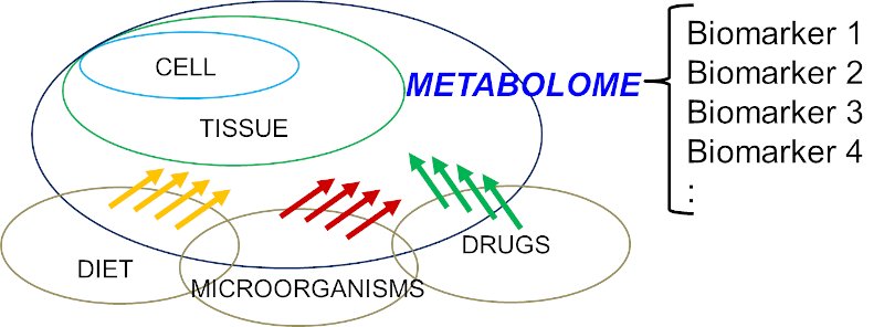
Knowledge of the human metabolomes and their metabolic interactions is important for the understanding of human diseases. For example, it is now recognized that mitochondria are not only a factory for oxidative phosphorylation and energy metabolism, mitochondria also orchestrated with over 1,000 proteins and linked with multiple biochemical processes [5]. Indeed, a disrupted mitochondrial homeostasis had also been observed in some organic acidurias [6, 7]. The interactions of human metabolomes are far more complex than once perceived and involve different cell types, diets, drugs, disease status, microorganisms, and many others (Figure 1). The collective ‘big picture’ can be better studied through exometabolomics [8-10]. For example, acetic acid and trimethylamine (TMA) have been identified as biomarkers of bacterial urinary tract infection (UTI) and Escherichia coli associated UTI, respectively [11, 12]. TMA is one of the examples of mammalian-microbial co-metabolism which the host metabolizes TMA into trimethylamine N-oxide (TMAO) via flavin monooxygenase 3 (FMO3), and in E-coli associated UTI, the endogenous TMAOs are converted back to TMA possibly through the action from bacterial TMAO reductase. Similar mammalian-microbial co-metabolism has been described in aspects of human health that include cardiovascular disease, immunity, gastrointestinal disorders, and cancer [13-16]. The whole metabolomics network is more complex when toxico-metabolomics from drugs, chemicals, and environmental pollutants are taken into account. To-date, 2,280 drug and drug metabolites have been reported in the DrugBank database, and 3,670 toxins and pollutants have been reported in the Toxin and Toxin Target Database [17, 18], and the databank is still expanding.


v16i2_fig1

Figure 1: Simplified diagram to illustrate the structure of a human metabolome which is a complex interplays between host (human) and various factors, e.g. diet, microorganisms, drugs, etc.


Metabolism is the heart of many disease processes. Insights gained from the knowledge of metabolism will inform diagnoses and lead to new treatments. For example, 116 treatable intellectual disability caused by IMD has been described in a 2021 review [19]. In this newsletter, more emphasis will be put on IMD, a heterogeneous condition involving disorders of synthesis, catabolism/anabolism, transport, and storage of metabolites.

Classifications of IMD

The definition for IMD is further refined as described in [20]. Indeed, the presence of an abnormal metabolite is no longer essential for the classification of IMD, but instead includes any condition resulting in the dysfunction of the specific enzymes or biochemical pathways that is intrinsic to the pathomechanism. In individuals, IMD is rare. However, in a population they are collectively “common”. The estimated incidence of IMD is 1 per 4,122 to 4,355 live births [21, 22]. The true prevalence of IMD is difficult to measure due to various factors. It was estimated as 1 in 800 to 2,500 newborns in one study in 2020 [23]. According to the Society for the Study of Inborn Errors of Metabolism (SSIEM), there are over 600 different IMDs, and they are grouped into 15 hierarchical classifications based on the biochemical pathway involved. They are (1) disorders of amino acids and peptide metabolism, (2) disorders of carbohydrate metabolism, (3) disorders of fatty acid and ketone body metabolism, (4) disorders of energy metabolism, (5) disorders of the metabolism of purines, pyrimidines and nucleotides, (6) disorders of the metabolism of sterols, (7) disorders of porphyrin and haem metabolism, (8) disorders of lipid and lipoprotein metabolism, (9) congenital disorders of glycosylation and other disorders of protein modification, (10) lysosomal disorders, (11) peroxisomal disorders, (12) disorders of neurotransmitter metabolism, (13) disorders of the metabolism of vitamins and (non-protein) cofactors, (14) disorders of the metabolism of trace elements and metals, and (15) disorders of and variants in the metabolism of xenobiotics (For more details, please refer to https://www.ssiem.org/resources/resources/inborn-errors-classification). This is a 2012 classification from SSIEM. Knowledge of IMDs is still expanding with the advancements in next-generation sequencing (NGS) which has led to the discovery of more disease-causing genes and disease classes in IMD. For example, new class, such as congenital disorders of autophagy, which cause multiple system involvement have been described in patients with inborn errors of neuro-metabolism [24].

Examples to enhance diagnostic workflow in IMD

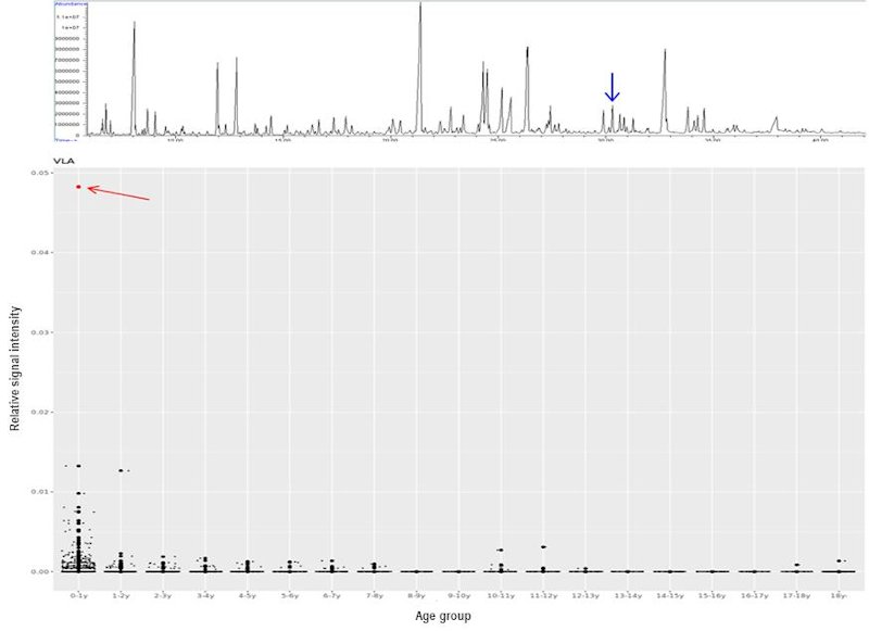
Recently, the International Classification of Inborn Metabolic Disorders assigned 1,450 monogenic conditions related to metabolism to 24 categories [25]. These conditions can have overlapping signs and symptoms. Some are rapidly fatal, mainly due to the accumulation of toxic metabolites and/or deprivation of energy; the diagnosis of IMD is clinically vital in this regard since it permits interventions to prevent further metabolic insults and irreversible damages. Unfortunately, there is no simple and single biochemical analysis that encompasses all pathognomonic markers in each IMD. Various methods had been described to decipher human metabolomes [26, 27]. Urine organic acid (UOA) via gas chromatography mass spectroscopy (GC-MS) was introduced in the 1960s and has since been adopted by most clinical laboratories. GC-MS is robust because it generates highly reproducible mass spectra, which allows positive identification using libraries, such as that of the National Institute of Standards and Technology (NIST). Nevertheless, there are several pitfalls of GC-MS. These include low-level metabolites, co-elution, age-dependent variation of metabolite levels, etc. Data interpretation by pathologists is a labour-intensive process. For this reason, an in-house automatic solution was established to address the above pitfalls and assist the UOA reporting process. A checklist composed of almost 100 key metabolites was constructed using over 1,600 sets of UOA GC-MS data and partitioned according to different age groups. Positive identification of metabolites was defined according to their retention times and electron ionisation spectra. The 95th percentile for each compound and in each age group was used as a cut-off to define abnormally high OAs, which would be flagged for in-depth review by chemical pathologists. This algorithm allows: (1) a graphical display of individual UOA levels and comparison with controls according to different age groups, (2) calculation of ratios of metabolites useful in interpreting low-level, but clinically significant, metabolites, (3) pathway analysis by a holistic correlation analysis of all studied OAs, and (4) continual database enrichment. An example of a case of aromatic L-amino acid decarboxylase (AADC) deficiency, a neurotransmitter disorder is shown in figure 2 where a significant increase of vanillactic acid (VLA) is identified. Despite this refinement, the analytical process is time consuming and remains a bottleneck for rapid diagnosis. To further streamline the analytical process for IMDs, we further explored the use of nuclear magnetic resonance (NMR) spectroscopy for IMD diagnosis as an alternative or complementary for GC-MS analysis. This approach for IMD was proposed decades ago in 1999 [28]. To-date, at least 100 IMD conditions can be diagnosed by NMR spectroscopy, as reviewed by Engelke [29] and Moolenaar [30]. In addition, this technique allows for the identification of novel IMDs. Examples include aminoacylase 1 deficiency in patients with metabolic brain diseases [31], beta-ureidopropionase deficiency in patients with movement disorders [32], defective polyol metabolism in patients with leukoencephalopathy [33], and dimethylglycine dehydrogenase in patients with muscle disorders [34].


v16i2_fig2

Figure 2: UOA spectrum by GC-MS from a case of aromatic L-amino acid decarboxylase (AADC) deficiency. The blue arrow points to the diagnostic marker, vanillactic acid (VLA) (Upper). Distribution of VLA levels in different age groups generated from in-house UOA algorithm. The AADC patient shows a marked increase of VLA (red arrow) comparing with age-matched subjects.


A national screening program in Turkey had applied NMR clinically to screen for IEM in newborns [35]. A total of 989 urine samples were collected from neonates and analysed by two laboratories. The results were used to establish a database and routine clinical screening. This NMR-based newborn urine screening has been further extended, covering up to 75 different IEM conditions [36]. The increasing awareness of clinical NMR applications has been further elaborated elsewhere in a 2021 review [37]. In our experience, the diagnostic utility of NMR has been substantiated in various clinical cases, for example, beta-ketothiolase deficiency, beta-ureidopropionase deficiency, citrin deficiency, fructose 1,6 bisphosphatase deficiency, holocarboxylase synthase deficiency, 3-hydroxyisobutyric aciduria, hyperornithinaemia-hyperammonaemia-homocitrullinuria (HHH) syndrome, methylmalonic aciduria (MMA), propionic acid, and succinic semialdehyde dehydrogenase deficiency (SSADHD). The merits of NMR-based urinalysis over GC-MS techniques are the simple sample preparation workflow and a relatively fast analytical time. Sample preparation is a two-step procedure that could be handled in

The choice of metabolic analysis depends on the nature of the pathognomonic metabolites. No single biochemical test that can detect them all. Pathologists have provided input concerning the choice of tests, advice on patient preparation and sample requirements, and clinical interpretations. The many examples include plasma acylcarnitine analysis, plasma/urine/CSF amino acids, urine acylglycines, bile acids, biotinidase activity, chitotriosidase activity, dried blood spot metabolic screening, homocysteine, transferrin isoelectric focusing, glycosaminoglycans analysis, CSF neurotransmitters, urine organic acids, urine sugars, urine guanidinoacetate and creatine analysis, urine oligosaccharides, orotic acids, red blood cells plasmalogens, porphyrins, phytosterols, pristanic and phytanic acids, purine and pyrimidines, very long chain fatty acids, and many more tests. Unfortunately, not all IMD-related tests are available or can be performed in a single centre.

The use of NGS will be a solution which provides additional insight from a genetic dimension, in particular if a biochemical assay is not available or the diagnosis cannot be easily demystified by biochemical tests. Primary coenzyme Q10 deficiency is one of the examples [38]. Affected individuals usually presented with non-specific symptoms and biochemical findings. Indeed, we have reported three cases of COQ4-related mitochondriopathy and identified a hotspot pathogenic variant in this locality using a genomics approach [39]. A plasma COQ10 assay was later developed for this potentially under-recognized condition.

Some IMD conditions are caused by multiple genes, for example, glutaric aciduria type II, methylmalonic aciduria (MMA), maple syrup urine disease (MSUD), propionic academia, phytosterolemia, congenital lactic acidosis, etc. Instead of a conventional gene-after-gene analysis, advances in NGS could allow the detection of the underlying genetic defect through a gene panel approach which effectively saves the time and manpower from managing a huge number of PCR primer bank and gene-specific protocols, not to mention the time spent in their revisions, updates and accreditations.

The diagnostic yield of NGS-based diagnosis for IMD is variable. In one report, NGS diagnosed 59% of the cases with clear clinical and biochemical features and a diagnostic yield of 8% for patients with an unclear phenotype [40]. Another group achieved an overall diagnostic yield of 50% and up to 78% in cases with a clear phenotype [41]. It is technically difficult to compare diagnostic yields of different studies, for reasons that include the scope of the IMD panel used, clinical and analytical aspects that can differ between centres.

Conclusions

With the expanding knowledge of metabolome and genome, more novel metabolites and genes have been discovered. These discoveries are enriching the understanding of IMD. Genomic and metabolomic analyses should be complementary to each other in the study of IMD, particularly in cases with atypical genetic findings or when a particular biochemical assay is not yet available. With the advancement of pharmacological chaperoning, small molecules and gene therapies, etc., more treatment options with improved care will be available in near future. At the same time, there will be increasing role from Pathologists for clinical use of cross-omics approach for disease diagnosis, monitoring and prognostication, with a more accurate and individualized characterization of disease progress.

Acknowledgement

The author would like to thank the supervision from Prof. Ching-wan Lam, Department of Pathology, The University of Hong Kong for his supervision on the NMR-related works. The author thanks Dr Gary Wong and Dr Jacky Ling, Division of Chemical Pathology, Department of Pathology, Queen Mary Hospital for their works on the in-house automatic solution in UOA analysis.


Reference

  1. Mussap, M., M. Zaffanello, and V. Fanos, Metabolomics: a challenge for detecting and monitoring inborn errors of metabolism. Ann Transl Med, 2018. 6(17): p. 338.
  2. Romero, P., et al., Computational prediction of human metabolic pathways from the complete human genome. Genome Biol, 2005. 6(1): p. R2.
  3. Sorokin, M., et al., Algorithmic Annotation of Functional Roles for Components of 3,044 Human Molecular Pathways. Front Genet, 2021. 12: p. 617059.
  4. Wishart, D.S., et al., HMDB 4.0: the human metabolome database for 2018. Nucleic Acids Res, 2018. 46(D1): p. D608-D617.
  5. Marchi, S., et al., MitopatHs: a new logically-framed tool for visualizing multiple mitochondrial pathways. iScience, 2021. 24(4): p. 102324.
  6. Chen, Z., M. Berquez, and A. Luciani, Mitochondria, mitophagy, and metabolic disease: towards assembling the puzzle. Cell Stress, 2020. 4(6): p. 147-150.
  7. Dimitrov, B., et al., Organic acidurias: Major gaps, new challenges, and a yet unfulfilled promise. J Inherit Metab Dis, 2021. 44(1): p. 9-21.
  8. Knott, M.E., et al., Metabolic Footprinting of a Clear Cell Renal Cell Carcinoma in Vitro Model for Human Kidney Cancer Detection. J Proteome Res, 2018. 17(11): p. 3877-3888.
  9. Lima, A.R., et al., Discrimination between the human prostate normal and cancer cell exometabolome by GC-MS. Sci Rep, 2018. 8(1): p. 5539.
  10. Palama, T.L., et al., Identification of bacterial species by untargeted NMR spectroscopy of the exo-metabolome. Analyst, 2016. 141(15): p. 4558-61.
  11. Lam, C.W., et al., NMR-based metabolomic urinalysis: a rapid screening test for urinary tract infection. Clin Chim Acta, 2014. 436: p. 217-23.
  12. Lam, C.W., et al., Quantitative metabolomics of urine for rapid etiological diagnosis of urinary tract infection: evaluation of a microbial-mammalian co-metabolite as a diagnostic biomarker. Clin Chim Acta, 2015. 438: p. 24-8.
  13. Claus, S.P., Mammalian-microbial cometabolism of L-carnitine in the context of atherosclerosis. Cell Metab, 2014. 20(5): p. 699-700.
  14. Brown, J.M. and S.L. Hazen, Microbial modulation of cardiovascular disease. Nat Rev Microbiol, 2018. 16(3): p. 171-181.
  15. Lin, L. and J. Zhang, Role of intestinal microbiota and metabolites on gut homeostasis and human diseases. BMC Immunol, 2017. 18(1): p. 2.
  16. Roager, H.M. and T.R. Licht, Microbial tryptophan catabolites in health and disease. Nat Commun, 2018. 9(1): p. 3294.
  17. Wishart, D.S., et al., DrugBank: a comprehensive resource for in silico drug discovery and exploration. Nucleic Acids Res, 2006. 34(Database issue): p. D668-72.
  18. Wishart, D., et al., T3DB: the toxic exposome database. Nucleic Acids Res, 2015. 43(Database issue): p. D928-34.
  19. Hoytema van Konijnenburg, E.M.M., et al., Treatable inherited metabolic disorders causing intellectual disability: 2021 review and digital app. Orphanet J Rare Dis, 2021. 16(1): p. 170.
  20. Morava, E., et al., Quo vadis: the re-definition of "inborn metabolic diseases". J Inherit Metab Dis, 2015. 38(6): p. 1003-6.
  21. Chong, S.C., et al., Expanded newborn metabolic screening programme in Hong Kong: a three-year journey. Hong Kong Med J, 2017. 23(5): p. 489-96.
  22. Lee, H.C., et al., Analysis of inborn errors of metabolism: disease spectrum for expanded newborn screening in Hong Kong. Chin Med J (Engl), 2011. 124(7): p. 983-9.
  23. Schuck, P.F., G.C. Ferreira, and M.C. McKenna, Recent advances in the pathophysiology of inherited metabolic diseases. Int J Dev Neurosci, 2020. 80(1): p. 50-51.
  24. Ebrahimi-Fakhari, D., et al., Congenital disorders of autophagy: an emerging novel class of inborn errors of neuro-metabolism. Brain, 2016. 139(Pt 2): p. 317-37.
  25. Ferreira, C.R., et al., An international classification of inherited metabolic disorders (ICIMD). J Inherit Metab Dis, 2021. 44(1): p. 164-177.
  26. Roca, M., et al., Reviewing the metabolome coverage provided by LC-MS: Focus on sample preparation and chromatography-A tutorial. Anal Chim Acta, 2021. 1147: p. 38-55.
  27. Tebani, A., C. Afonso, and S. Bekri, Advances in metabolome information retrieval: turning chemistry into biology. Part I: analytical chemistry of the metabolome. J Inherit Metab Dis, 2018. 41(3): p. 379-391.
  28. Bamforth, F.J., et al., Diagnosis of inborn errors of metabolism using 1H NMR spectroscopic analysis of urine. J Inherit Metab Dis, 1999. 22(3): p. 297-301.
  29. Engelke, U., et al., Handbook of 1H-NMR spectroscopy in inborn errors of metabolism: body fluid NMR spectroscopy and in vivo MR spectroscopy: SPS Publications, 2007. 2007.
  30. Moolenaar, S.H., U.F. Engelke, and R.A. Wevers, Proton nuclear magnetic resonance spectroscopy of body fluids in the field of inborn errors of metabolism. Ann Clin Biochem, 2003. 40(Pt 1): p. 16-24.
  31. Engelke, U.F., et al., NMR spectroscopy of aminoacylase 1 deficiency, a novel inborn error of metabolism. NMR Biomed, 2008. 21(2): p. 138-47.
  32. Moolenaar, S.H., et al., beta-Ureidopropionase deficiency: a novel inborn error of metabolism discovered using NMR spectroscopy on urine. Magn Reson Med, 2001. 46(5): p. 1014-7.
  33. Moolenaar, S.H., et al., In vivo and in vitro NMR spectroscopy reveal a putative novel inborn error involving polyol metabolism. NMR Biomed, 2001. 14(3): p. 167-76.
  34. Moolenaar, S.H., et al., Defect in dimethylglycine dehydrogenase, a new inborn error of metabolism: NMR spectroscopy study. Clin Chem, 1999. 45(4): p. 459-64.
  35. Aygen, S., et al., NMR-Based Screening for Inborn Errors of Metabolism: Initial Results from a Study on Turkish Neonates. JIMD Rep, 2014. 16: p. 101-11.
  36. > Embade, N., et al., NMR-based newborn urine screening for optimized detection of inherited errors of metabolism. Sci Rep, 2019. 9(1): p. 13067.
  37. Speyer, C.B. and J.D. Baleja, Use of nuclear magnetic resonance spectroscopy in diagnosis of inborn errors of metabolism. Emerg Top Life Sci, 2021.
  38. Yubero, D. and R. Artuch, NGS for Metabolic Disease Diagnosis. EJIFCC, 2018. 29(3): p. 227-229.
  39. Ling, T.K., et al., Clinical whole-exome sequencing reveals a common pathogenic variant in patients with CoQ10 deficiency: An underdiagnosed cause of mitochondriopathy. Clin Chim Acta, 2019. 497: p. 88-94.
  40. Ghosh, A., et al., Diagnosing childhood-onset inborn errors of metabolism by next-generation sequencing. Arch Dis Child, 2017. 102(11): p. 1019-1029.
  41. Yubero, D., et al., Targeted Next Generation Sequencing in Patients with Inborn Errors of Metabolism. PLoS One, 2016. 11(5): p. e0156359.
Single Tags: 

Diagnosis of COVID-19

Diagnosis of COVID-19


Volume 16, Issue 1, January 2021  (download full article in pdf)


Editorial note


Coronavirus disease 2019 (COVID-19) is undoubtedly the most topical subject not only in the medical field, but also for humanity globally. In this issue of the Topical Update, Dr. Derek Hung and Prof. Kwok Yung Yuen present an overview on the diagnosis of COVID-19, which underpins effective disease control. We welcome any feedback or suggestion. Please direct them to Dr. Janice Lo, Education Committee, The Hong Kong College of Pathologists. Opinions expressed are those of the authors or named individuals, and are not necessarily those of the Hong Kong College of Pathologists.


Dr. Derek HUNG and Prof. Kwok Yung YUEN

Resident, Department of Microbiology, Queen Mary Hospital, Hospital Authority and

Professor, Department of Microbiology, Faculty of Medicine, The University of Hong Kong


Overview

Coronavirus disease 2019 (COVID-19) caused by severe acute respiratory syndrome coronavirus 2 (SARS-CoV-2) since December 2019 has infected 54 million population in all six major continents, resulting in over 1.3 million deaths by mid-November 2020. One of the most important aspects in curbing the spread of the virus is rapid yet accurate diagnosis of infection followed by timely isolation and contact tracing. Molecular testing is now the mainstay of diagnosis, supplemented by viral antigen testing. Antibody detection aids in assessment of immunity and disease prevalence in the population. As the disease progresses, there are worldwide efforts in developing a multitude of diagnostic platforms, both in-house and commercial. Studies also endeavour to assess optimal types and timing of specimen collection to enhance diagnostic yield. In this review, we would look at some of the knowledge and practices in making a diagnosis of COVID-19.

Specimen collection

Obtaining the best specimen optimizes the possibility of getting the correct diagnosis based on clinical suspicion. Being a predominantly respiratory pathogen, obtaining respiratory specimens for viral detection remains the primary modality for making a diagnosis of acute infection by SARS-CoV-2. The viral load is highest at or soon after symptom onset, with the viral load in the upper respiratory tract peaking earlier than the lower respiratory tract. The viral load decreases in the respiratory tract at a rate of 1 log10 per week. The World Health Organization (WHO) suggests that testing upper respiratory tract specimens is adequate for early stage infection, especially asymptomatic or mild cases. The Centers for Disease Control and Prevention (CDC) recognizes nasopharyngeal swab, nasopharyngeal wash, nasal wash obtained by health care professionals; nasal mid-turbinate swab, nasal swab obtained by either health care professionals or supervised self-collection on site; and posterior oropharyngeal saliva (POS) by supervised self-collection as valid specimens. Patients with lower respiratory tract symptoms such as productive cough, shortness of breath, or suspicious radiological findings should send sputum to enhance sensitivity. Induced sputum is not recommended due to increased risk of aerosol transmission,. Among different respiratory specimens, broncho-alveolar lavage (BAL) showed the highest positive rate.

For the upper respiratory tract specimen, comparing combined nasal swab/throat swab with nasopharyngeal swab, Vlek et al showed high concordance between these two methods (kappa coefficient 0.95) despite the cycle threshold value (Ct value) obtained from nasopharyngeal swab being lower. Another study suggested nasal swab alone also has good concordance with nasopharyngeal sampling. In contrast, oropharyngeal swab alone has inferior performance. Wang et al showed the sensitivity of oropharyngeal swab was 21.1% and meta-analysis by Bwire et al suggested the positive rate is as low as 7.6% in suspected cases, comparing with 69.6% and 71.3% for nasopharyngeal swab and lower respiratory tract specimen respectively. POS is increasingly studied as an alternative respiratory tract specimen for diagnosis. Theoretically well produced POS can concentrate secretions dripping down from nasopharynx and lower respiratory secretion moved up by ciliary activity of respiratory epithelium. It can be saved by patients themselves with instructions, thus reducing discomfort in specimen collection and minimizing aerosol exposure for health care professionals. The cost of collecting POS could be 2.59-fold lower than nasopharyngeal specimen, which could be significant in resource limited setting. The concordance between POS and nasopharyngeal swab is high, especially in the first 7 days of infection, up to 96.6% positive percent agreement. The sensitivity is comparable with nasopharyngeal swab in properly collected specimen. The sensitivity does not vary much between early morning and at least 2 hours after meal, which provides a convenient option for specimen collection. CDC and Hospital Authority of Hong Kong have adopted POS as an alternative option for upper respiratory specimen collection.

Viral shedding is also found in other specimens with stool being more studied. Meta-analysis showed viral shedding was found in faecal material in 40.5% of patients. The viral shedding in stool is more prevalent in those with gastrointestinal symptoms and may last longer than the shedding in respiratory tract. Viral RNA detected in blood and urine is relatively uncommon, respectively only 1% and 0% in one study with more than 200 patients10. Even without ocular symptoms, the conjunctival secretion may contain a small amount of SARS-CoV-2 RNA in around 8% of patients, warranting appropriate infection control measure in ophthalmological assessment.

Molecular testing

Detection of nucleic acid remains the backbone of diagnosing COVID-19 for treatment and public health purposes. Reverse-transcriptase polymerase chain reaction (RT-PCR) is the most widely used technique. After transcribing the viral RNA into complementary DNA (cDNA) with reverse transcriptase, the cDNA would be amplified and detected by real-time PCR. Potential molecular targets for SARS-CoV-2 include genes encoding structural proteins, e.g. spike (S), envelop (E), helicase (hel), nucleocapsid (N-N1 and N2), transmembrane (M); and non-structural regions, e.g. RNA-dependent RNA polymerase region (RdRp), haemagglutinin-esterase (HE), and open reading frame 1a (ORF1a) and ORF1b. Most scientific institutes and commercial platforms would design primers to target more than one gene, or to target multiple loci of the same gene to enhance diagnostic sensitivity and specificity. Though N gene RNA is shown by nanopore direct RNA sequencing study to be the most abundantly expressed transcript in SARS-CoV-2 infected cells, there is no consensus on which gene confers the best diagnostic performance. Presently, one conserved and one specific target region are recommended to mitigate effect of random mutation or genetic drift while maintaining specificity25. Various regimens for testing are proposed in the literature. Corman et al recommended the Charité protocol, which was to use E gene for screening and RdRp gene for confirmation. CDC used N1 and N2 genes as their diagnostic panel. Chu et al used N gene as screening test and ORF1b as confirmatory assay because the screening N gene assay is 10 times more sensitive than ORF1b. As an alternative confirmatory assay, Chan et al developed a real-time RT-PCR assay locally, targeting RdRp/Hel. This COVID-19-RdRp/Hel assay demonstrated significantly higher sensitivity and specificity for the detection of SARS-CoV-2 RNA than the RdRp-P2 assay in clinical evaluation.

Multiple commercial platforms were developed for molecular SARS-CoV-2 diagnosis for their high throughput, rapid turnaround time and ease of use with automation. Examples are Roche Cobas 6800/8800 system (targets ORF1a and E genes) and Abbott Alinity m SARS-CoV-2 assay (targets RdRp and N genes), where sample preparation, genetic material extraction, target amplification and result reporting are automated inside the system. Molecular point-of-care testing (POCT) refers to diagnostic platform that is portable (often desktop-size), requires minimal sample preparation steps and can provide reliable molecular results within 2 hours. POCT like Cepheid GeneXpert (Xpert Xpress SARS-CoV-2 assay, targets E and N2 genes) enables rapid testing near the site of collection in areas with little laboratory support. Fewer steps in manipulation reduce risk of cross contamination and laboratory error in processing. Many evaluation studies have been published to compare the performance of these commercial platforms against in-house diagnostic tests and for head-to-head comparison between platforms. For example, Cobas system is shown to have high diagnostic agreement with in-house molecular assays,, as well as with other commercial platforms such as Hologic Panther Fusion system and Cepheid GeneXpert. Cepheid GeneXpert reaches an agreement of 100 % compared to three in-house RT-PCRs in a multicentre evaluation in the Netherlands. Among commercial platforms there might be minor discordance between assays at very high Ct values and the viral load of clinical samples used in evaluative studies should be noted in particular.

Another molecular technique is reverse-transcriptase loop-mediated isothermal amplification (RT-LAMP) test. Using multiple primers for the genetic target, RT-LAMP amplified nucleic acid by strand displacement in an isothermal condition of around 60- 65oC. It allows synthesis of large amount of genetic material up to 106 to 109 copies of target DNA within 30-60 minutes2. Without the need of thermal cycler as in RT-PCR, RT-LAMP facilitates development of rapid molecular POCT and has an expanding market in commercial diagnostic platform. On the down side, since multiple primers over a relatively small genetic region are needed for amplification, there are constraints in properly designing the primers. Abbott ID NOW is a commercial POCT platform using RT-LAMP, allowing real time detection of SARS-CoV-2 within 15 minutes targeting RdRp gene. Evaluation of ID NOW against other RT-PCR based platforms appears suboptimal in terms of diagnostic sensitivity. Compared to Cobas, ID NOW achieved only 73.9% positive agreement while GeneXpert achieved 98.9% positive agreement. In samples with Ct values greater than 30, positive agreement was 34.3% for ID Now and 97.1% for GeneXpert. A lower sensitivity of ID NOW over GeneXpert was also reported in another evaluation by Basu et al. In contrary, good diagnostic utility has been demonstrated in many other centres including Hong Kong that have designed their own RT-LAMP for COVID-19. Chow et al reported sensitivity of 95% at 60 minutes using RT-LAMP targeting a region across ORF3a/E gene as compared to RT-PCR. Lu et al achieved concordance rate of 93% against RT-PCR using in-house E gene RT-LAMP assay.

In order to improve the diagnostic sensitivity of molecular assays, clustered regularly interspaced short palindromic repeat (CRISPR)-based technology has been employed by coupling with Cas enzyme. The enzyme would be directed to the target DNA/RNA by a guide RNA complementary to the target sequence. Once bound, the collateral nuclease activity of the Cas enzyme would cleave surrounding reporter fluorophore and lead to signal amplification. DETECTR technology uses Cas12a enzyme to bind target DNA; while SHERLOCK technology uses Cas13a enzymes to bind target RNA. This technology can be incorporated in molecular techniques especially RT-LAMP to enhance the sensitivity and to lower the detection limit.

Next generation sequencing (NGS) enables sequencing of the entire genome in a relatively short period of time. Sharing of genetic data facilitates tracking of disease spread, understanding of disease transmission route, monitoring viral genome evolution and detecting emergence of mutation that may escape detection or enhance virulence. The cost and infrastructure required of NGS and the need of bioinformatics expertise limit its use to larger hospital and research centres.

Antigen detection

Like other respiratory viruses such as influenza and respiratory syncytial virus (RSV), direct antigen detection from respiratory specimen especially nasopharyngeal sample is another way of making a diagnosis of COVID-19. N protein was found previously to be the predominant structural protein released in large amount in nasopharyngeal aspirate during infection of SARS-CoV, and the same phenomenon is also shown in SARS-CoV-2 where the abundantly expressed N protein is widely used as an antigen detection target in COVID-19. Detection is achieved by capturing viral antigen in clinical specimens by monoclonal antibodies or monospecific polyclonal antibody fixed on a membrane, usually indicated by colour change of the strip in colorimetric lateral flow immunoassay. The assay can be delivered as POCT in an office setting since no complex laboratory support is required and the result can be available within a short period of time, usually <30 minutes. The major setback is the suboptimal sensitivity as compared to molecular diagnosis especially in samples with high Ct values. Evaluation by Lambert-Niclot et al using COVID-19 Ag Respi-Strip CORIS, a nitrocellulose membrane technology with colloidal gold nanoparticles sensitized with monoclonal antibodies directed against SARS-CoV-2 nucleoprotein (NP) antigens, showed sensitivity of only 50% when compared against multiple RT-PCR platforms. For samples with Ct value <25, the sensitivity is higher at 82.2%. In a local evaluation using Biocredit COVID-19 Ag test, the antigen test is 105 fold less sensitive than RT-PCR and it yielded a positive result in 45.7% RT-PCR positive combined nasopharyngeal swab/throat swab specimens only. There are attempts to improve sensitivity of rapid antigen assay. Porte et al evaluated an immune-chromatographic antigen assay using fluorescence signal showing sensitivity of 93% but the Ct value of the sample included in this study is relatively low with mean of 20. Other approaches by concentrating the antigen in specimens before testing with monoclonal antibodies targeting multiple different epitopes of the antigen were also reported. Based on a meta-analysis by Dinnes et al, the average sensitivity is around 56.2% for antigen assay with a high average specificity of 99.5%. Further refinement in antigen detection employs the detection of the change in bioelectric property by antigen binding to the antibody coated membrane. In Seo et al, anti-S antibody binds to SARS-CoV-2 particles to fabricate graphene-based field-effect-transistors (FET) biosensors and can respond down to 16 pfu/mL of virus. One challenge to this advance is the high background noise which can reduce sensitivity of detection. Overall, rapid antigen detection serves only an adjunctive role to molecular assay in making a diagnosis especially in outbreak situation where prevalence is high and molecular assay is not available. WHO has issued interim guidance of use of rapid antigen immunoassays.

Antibody detection

While antibody testing may not be useful in acute setting for COVID-19, it helps establish retrospective diagnosis, predict immunity and understand seroprevalence in a defined community. Commonly employed techniques are lateral flow immunoassay, chemiluminescent immunoassay, immunofluorescent assay, and enzyme-linked immunosorbent assay (ELISA). Median seroconversion times following symptom onset are 11 days for total antibodies, 12 days and 14 days for IgM and IgG respectively. Detection rate for IgM ranges from 11-71% in the first 7 days of infection, 36-87% between 8-14 days, and 56-97% after 14 days. For IgG, it ranges from 4-57% in first 7 days, 54-88% between 8-14 days, and 91-100% after 14 days. For SARS-CoV-2, there does not seem to have significant time difference between IgM and IgG response. IgM peaked at around 3 weeks after symptom onset and fell to baseline level at around day 36. The duration of IgG seropositivity remains unknown and longer longitudinal studies are required. Study from Iceland involving over 1200 confirmed patients showed no evidence of antiviral antibody decline by 4 months after diagnosis; and most other studies showed persistently detectable antibodies by 2-3 months after infection60. On the other hand, there are some evidences that the IgG level may decline faster in mild and asymptomatic61 COVID-19 cases.

S protein is an important antigen for neutralizing antibody production. The S1 domain is responsible for receptor binding while the S2 domain is responsible for fusion. The receptor binding domain (RBD) is located at S1. NP, which is a structural component of the helical nucleocapsid, also appears to be an important antigen for the development of serological assays to detect COVID-19. Earlier in the pandemic, using sera collected more than 14 days after symptom onset from 16 patients, To et al showed rates of seropositivity were 94% for anti-NP IgG, 88% for anti-NP IgM, 100% for anti-RBD IgG, and 94% for anti-RBD IgM. Another study compares sensitivity and specificity in testing anti-S and anti-NP IgG for evidence of immunity across multiple platforms, which shows they are comparable by day 37 after infection though seroconversion of anti-NP IgG may precede anti-S IgG by around 2 days (day 9-10 v day 11-12). Caruana et al observed that the decline of anti-NP antibody may be faster than anti-S and thus could be less sensitive longer after infection. Also titre of anti-S antibody may better reflect protection against reinfection67. Multiple commercial platforms were developed for high-throughput antibody testing in clinical laboratory. Automatic platforms such as Abbott SARS-CoV-2 IgG, which is a chemiluminescent micro-particle immunoassay, are also used in public hospital of Hong Kong for a shorter turnaround time.

Neutralization antibody test is important in assessing in vitro the functional capacity of the humoral response of COVID-19 patients to prevent reinfection by the virus. Traditional neutralization assay such as microneutralization and plaque reduction assay require manipulation of live virus and necessitate biosafety level 3 laboratories. As a result, pseudovirus neutralization assay has been developed. Vesicular stomatitis virus (VSV) expressing S protein of SARS-CoV-2, containing the RBD, is used so that the assay can be performed in biosafety level 2 facilities. SARS-CoV-2 neutralizing antibody starts to rise at around 7-10 days after symptom onset and the median peak time is 33 days after symptom onset. The neutralization titres then decline in 93% of the patients and by a median level of 35% over 3 months. Patients with more severe disease requiring ICU admission have accelerated and augmented neutralizing antibody response compared with non-ICU cases. In non-severe cases who have low peak neutralizing antibody titre, neutralizing antibody level might return to baseline within 2 months. Another clinical use of neutralization assay would be to confirm potentially false positive SARS-CoV-2 serology result. Three children with Kawasaki disease without symptoms or epidemiological linkage to COVID-19 were tested positive to anti-RBD and anti-NP antibodies by a microparticle-based immunoassay but were confirmed negative by microneutralization test.

Studies have shown there are serological cross-reactivity between SARS-CoV-2 and SARS-CoV. Testing sera taken from COVID-19 patients by ELISA, cross-reactivity is seen against S protein and RBD of SARS-CoV, though the intensity of cross-reaction against RBD is weaker than S protein. For the full length S protein, the amino acid sequence homology between SARS-CoV-2 and SARS-CoV is around 75%. The homology between them for RBD which is located in S1 domain is around 74%. For the receptor binding motif (RBM) of the RBD where the virus directly binds to angiotensin-converting enzyme 2 (ACE2), the homology is only 50%. The degree of amino acid homology explains the difference in the level of cross-reaction between them on ELISA. Chia et al showed even more significant cross-reactivity between SARS-CoV-2 and SARS-CoV antibody against NP by Luminex assay than antibody against S1 or RBD as the homology between the NP of these 2 viruses is around 90%. Despite some cross-reaction between antibodies against RBD on ELISA, there does not seem to have significant cross neutralization effect73. Only 1 out of 15 COVID-19 sera showed cross neutralization with SARS-CoV at very low titre. Overall the effect of cross-protection in vaccination and whether antibody-dependent enhancement effect would be seen between these 2 closely related viruses remains unknown.

Cross-reactivity against other human coronaviruses in SARS-CoV-2 infection has been investigated in a few trials. In a study by Wölfel et al, using immunofluorescence assay against recombinant S protein, cross-reactivity of SARS-CoV-2 sera is found against human coronaviruses OC43, NL63, HKU1 and 229E on comparing the titres between admission and convalescence samples, especially HKU1 and OC43 which are both betacoronavirus. In Shrock et al, deep serological profiling of sera from SARS-CoV-2 patients and pre-COVID sera are performed. Antibodies against S and NP are the most specific assay to differentiate SARS-CoV-2 and pre-COVID sera. Those with dramatic increase in anti-S antibody after COVID-19 infection also have increase in the intensity of cross-reactivity against other human coronaviruses, especially over more homologous regions of the S protein e.g. at residue 811-830 and 1144-1163. It could be novel antibodies of SARS-CoV-2 that cross-react or boost the anamnestic response against SARS-CoV-2 infection due to existing memory towards other human coronaviruses from past exposure. Moreover, pre-COVID sera also show some cross-reaction towards the homologous region of SARS-CoV-2 S protein and ORF1 in the same study.

Viral culture

Demonstration of live SARS-CoV-2 in cell culture requires biosafety level 3 facilities and are not routinely performed in most of the clinical laboratories. However, live virus isolation is still important for some diagnostic and research purposes so as to determine whether the amount of virus present is infectious to others, to evaluate therapeutic efficacy of potential antiviral compound, to develop viral neutralization assay for testing convalescent sera, to provide positive control for molecular assay development, and to develop vaccine strains. The host cell receptor for SARS-CoV-2 is ACE2. Non-human cell lines such as Vero E6 and Vero CCL-61 which have abundant ACE2 expression are commonly used for isolation. Cytopathic effect is seen by 3 days after inoculation. SARS-CoV-2 also grows in human continuous cell lines such as Calu3 (pulmonary cell line), Caco2 (intestinal cell line), Huh7 (hepatic cell line), and 293T (renal cell line). It grows modestly on U251 (neuronal cell line) which is not seen in SARS-CoV81. Confirmation of SARS-CoV-2 replication in the cell line can be done by molecular testing or immunostaining techniques. Cell lines can be engineered to express a transmembrane serine protease TMPRSS2 for priming of S protein and to facilitate the entry of SARS-CoV-2 into host cell. Organoid systems such as bat and human intestinal organoids are susceptible to SARS-CoV-2 and are developed to better study tissue tropism, the dynamics of infection and testing of therapeutic targets.

Radiological diagnosis and artificial intelligence

There are no pathognomonic radiological features on chest imaging for COVID-19 and the disease should not be ruled in or ruled out based on imaging alone. However, presence of suggestive imaging features can prompt further investigations in suspicious cases, such as lower respiratory tract viral testing for confirmation. Reports in literature have suggested that in some patients, radiological findings may precede the detection of SARS-CoV-2 in clinical specimen,. Chest X-ray (CXR) is a less sensitive modality than computed tomography of the thorax (CT thorax) with a reported CXR sensitivity of 69%85. As in other viral pneumonia, COVID-19 typically presents with multifocal air-space disease, especially with a bilateral lower lung distribution. More specific to COVID-19, it tends to have peripheral lung involvement, seen in 58% of CXR in one study. CT thorax has a higher sensitivity than CXR, quoted at around 60-98%. CT thorax often demonstrates the typical findings of peripheral bilateral ground glass opacities (GGO) with or without consolidation or ‘crazy-paving pattern’. Sometimes the GGO would arrange in a rounded pattern. Isolated lobar or segmental consolidation without GGO, centrilobular shadows, cavitory changes, lymphadenopathy and pleural effusions are rare86. As the disease advances, the opacities might coalesce, affecting central and bilateral upper lobes and may manifest as ‘white lung’ with diffuse infiltrate. The abnormalities usually peak by 2 weeks after symptom onset, replaced by scar tissue with recovery. In the COVID-19 pandemic, artificial intelligence (AI) programme is increasingly studied for screening abnormal radiological result which would be particularly useful for mass screening strategy in outbreak situation. The performance of AI is dependent on the radiological imaging algorithm being fed into the system for deep learning process. So far the result of this research has been promising with reported area under receiver operating characteristic curves greater than 0.9,. However, there are still lots of technical and ethical issue to resolve which include dataset bias, data privacy, and the distribution of ultimate accountability of result.

Detection of host inflammatory reaction

In COVID-19, there are studies to diagnose and predict severe diseases by the host inflammatory response. Apart from direct viral damage, uncontrolled cytokine storm triggered by the virus leads to tissue damage and multiorgan failure. Mean interleukin-6 (IL-6) concentration in serum was found to be 2.9 fold higher in patients with complicated COVID-19 disease than non-complicated disease. It became one of the markers clinicians could use to predict progression into severe disease. Roche Elecsys IL-6 immunoassay received FDA Emergency Use Authorization to help identify patients at high risk of requiring intubation with mechanical ventilation. Molecules targeting IL-6 such as tocilizumab are also studied as therapeutic to prevent disease progress by blocking the inflammatory pathway. It does not show efficacy in preventing intubation or death in moderately ill hospitalized patients in the BACC Bay trial. Elevated CRP is associated with worse outcome, as well as elevated IL-10 which may be related to compensatory anti-inflammatory response and secondary infections. Haematologically, severe disease is associated with higher absolute neutrophil count, D-dimer and LDH but lower absolute lymphocyte101 and platelet count.

Conclusion

Global COVID-19 pandemic stimulates global effort in development of rapid yet accurate diagnostic techniques. Diagnosis is often limited by the low level of viral particles in the specimen and the subtle clinical features in early infection. Though traditional methods like RT-PCR are still the mainstay, we see expanding endeavours to strive for higher speed and lower limit of detection at an earlier time. Molecular techniques such as RT-LAMP, CRISPR/Cas, biosensor technology in antigen detection, AI operating system for image interpretation are pushing the diagnostic ability to the limit. Despite these scientific advances, there are still a lot of gaps to fill especially in understanding the nature and duration of humoral immunity response and its protection against re-infection. All these require continuous global cooperation and information exchange to make them possible.


Single Tags: 

Drowning: A Rational Approach to its Diagnosis

Drowning: A Rational Approach to its Diagnosis


Volume 15, Issue 2, July 2020  (download full article in pdf)


Editorial note:


Drowning often presents in various scenarios depending on the circumstances. This Topical Update provides a proper approach to the diagnosis. We welcome any feedback or suggestions. Please direct them to Dr. FOO Ka-chung of Education Committee, the Hong Kong College of Pathologists. Opinions expressed are those of the authors or named individuals, and are not necessarily those of the Hong Kong College of Pathologists.


Dr. FOO Ka-chung

Specialty Coordinator (Forensic Pathology), Education Committee

The Hong Kong College of Pathologists


Introduction

Drowning is referred as “death occurring within 24 hours of a submersion incident”. Definition by World Health Organization is "the process of experiencing respiratory impairment from submersion or immersion in liquid ". [1,2] It is a form of asphyxia with a distinct pathophysiology and mechanism of death. It is also a diagnosis by exclusion, and therefore every piece of information should be regarded as crucial. Pathologists are obliged to work under Coroner’s jurisdiction in interviewing the next-of-kin (if available), reviewing antemortem medical records and preliminary findings provided by investigating officer, performing an autopsy as directed and compiling reports capable of addressing anticipated issues.

As Forensic Pathologists mostly deal with sudden and unexpected deaths, cases of drowning with unsalvageable outcome are often encountered. Hospital Pathologists, on the other hand, are dealing with patients presenting a clinical picture in which death eventually occurred after vigorous cardiopulmonary resuscitation followed by development of various systemic complications, e.g. pneumonia, acute respiratory distress syndrome, multi-organ failure, disseminated intravascular coagulopathy, hypoxic-ischaemic encephalopathy etc.


Manner of Death

Information derived from the Coroner’s Report [3] and the Centre for Health Protection [4] suggested that majority of cases were accidental or suicidal in nature. Only a few were homicides. However, it should be remembered that a body found immersed in water does not necessarily imply a diagnosis of drowning. Nor its manner be automatically presumed basing on the prevalent trend. The deceased can die of natural conditions preceding or during submersion as well as unnatural elements that contributed to the drowning process, explaining the failure of extrication from water versus genuine lethal trauma before or while in water. [5,6]


Pathophysiology

The mechanism of death is complex involving changes to viscera, biochemical alterations and also at a cellular level. The culprit is the medium imposing hydrostatic and osmotic effect to the lungs. [7] The acute change in intravascular volume with electrolyte imbalance is the consequence. Several stages of drowning present in response to the rising levels of carbon dioxide and decreasing oxygen tension in blood. Voluntary breath holding for about 1 to 2 minutes is followed by a stage of involuntary urge to breath with aspiration of fluid for about 1 to 3 minutes. Tonic-clonic seizures, together with some degree of respiratory activity, will occur in the next 1.5 minutes with eventual involuntary breath holding and terminal gasping before cessation of cardiac activity. [1]

It has even been mentioned that only a few inches of water is sufficient to drown a person, as in the case of sudden incapacitation by onset of acute illness while standing close to a washbasin or bucket. [5,6] It was reported that about 1 mL/kg to 11 mL/kg of water aspirated can result in drowning. [7]

A rare entity underdiagnosed in daily practice, or seldom made by pathologists, is referred as "dry drowning" or "immersion syndrome", with negative autopsy findings of typically drowned lungs due to severe laryngeal spasm, therefore preventing further intake while stimulating the sensitive receptors and subsequently triggering cardio-inhibitory reflexes (Ebbecke reflex, Aschner reflex, Hering reflex). [1,5,6,7,8]


Diagnosis of Drowning

The possibility of drowning should always be considered when a deceased was recovered from a body of fluid or the head was found submerged inside a medium of fluid. The deceased could be found near a body of fluid where it could be washed onto the rocky shore, beach, or riverbank. Domestic environments such as bathtub also house this potential danger affecting all walks of life, especially for those who have chronic illness with sudden unexpected precipitation or the young. While the diagnosis of drowning could be straightforward one, such as a witnessed fall into water with subsequent submersion, it can be extremely difficult when critical information derived from the case is absent or inconclusive. Challenging scenarios can appear with unclear circumstances preventing proper formulation of the manner of death. Moreover, while findings derived from postmortem and ancillary investigations may collaborate with the diagnosis, it can be equally confusing when concomitant conditions are unveiled.

Presence of a natural condition which may contribute to death

Let’s consider the following case:

A 51 year-old female was found collapsed underwater in a public swimming pool of about 1.4 meters deep and was certified dead despite intensive resuscitation. There was no eye witness leading to her collapse. Autopsy revealed severe ischaemic heart disease with no evidence of acute infarction. Both lungs were congested and oedematous but frothy fluid was absent probably due to suction during resuscitation. Cause of death is labelled as drowning as the overall features were compatible with drowning.

The presence of a co-existing medical condition, be it undiagnosed or known to the deceased, has to be evaluated carefully to attribute its extent of contribution to death. A sudden precipitation into cardiac arrhythmia explained the reason why a habitual swimmer is incapacitated and eventually succumbed in the water. From the investigator’s point of view, possible legal issues regarding adequate supervision of the swimming environment may be raised which could lead to possible lawsuit and inquest. As such, the pathologist should be ready to address the extent of contribution of medical condition to the tragic outcome.

Medical background of the deceased has to be thoroughly reviewed including conditions such as asthma, epilepsy, cardiovascular conditions (e.g. Long QT syndrome type 1). Psychiatric history including substance abuse should also be elicited.

Presence of trauma which may be related to death

Another case is presented here:

An 87 year-old female was found floating off shore from a pier. She was known to be a habitual swimmer and there was no known chronic illness. There were multiple lacerations on chest and right upper limb. The thoracic cavity was breached and right lung had collapsed. Tinge of frothy fluid was noticed briefly by paramedics before transportation to mortuary. Autopsy revealed severe coronary stenosis and the left lung was mildly hyperinflated.

The presence of trauma may or may not be related to death as injuries inflicted can be produced ante-mortem or post-mortem. Assessment for vital reactions at the wound margin may be helpful to determine its nature.

All forms of injuries must be explained correlating inanimate objects in the environment. Sliding abrasions may be inflicted upon skidding down a slope while blunt force injuries may be a genuine assault. Self-inflicted injuries may occur in suicide as a back-up technique, for example, a stab to the chest or incised wound on the neck, yet it might at times mimic a homicide.

Dragging effects as a result of contact with river bed or ocean floor propelled by sea waves or tidal current is not uncommon and should be interpreted in light of such movement in water. Abrasions or lacerations may be found on forehead, dorsum, knees and toes. In addition, aquatic animal activity, such as crustaceans, will produce bites and nipping around orifices. The body, on the other hand, may be struck by watercraft or its parts including the propeller, predominantly located below the waist and over the extremities while the subject is maintaining a vertical position. It should be located posteriorly upon floating postmortem. [5,6] At times injuries could be severe enough to hinder the diagnosis by producing serious disruption of the viscera. The presence of postmortem mutilation further complicate the diagnosis, let alone in jeopardizing the facial features and hindering identity as often encountered in mass fatalities.

Healthy adults who can swim rarely drown unless there is an intervening reason such as superimposed injury, fatigue or dangerous environment. The level of fitness, history of risk-taking behaviour, pre-swim activities, swimming ability and experience should be explored.

In the present case, the cause of death is labelled as drowning and suggested an accidental manner with sudden precipitation of undiagnosed cardiac condition, complicated by postmortem propeller injuries by marine traffic, evidenced by lack of blood infiltration at the site of traumatic amputation.

Let’s consider another case:

An 80 year old male, who was an inmate of old aged home with multiple comorbidities confined to a wheelchair, was found submerged underneath river. He was last seen swaying around a footbridge about 3 meters above the river several hours earlier. Probable suicidal intention was identified. Autopsy revealed extensive comminuted fractures of the vault, subarachnoid haemorrhages and cortical contusions. Both lungs did not appear to be waterlogged.

Injuries may also be produced before or upon entering water and their extent have to be assessed. This could be related to subsequent question of survivability. In this case, considering the severity of the head injuries, it would appear that the deceased was unable to survive in water (or at most only a transient period) and succumbed rapidly. The cause of death is therefore attributed to head injuries upon falling with his top of head bumping the river bed.

Another case to ponder:

A 33 year-old female was found submerged about 20 meters off shore. Linear reddish bruising was found on the anterior neck. The face and eyes were congested with petechiae. Small amount of frothy fluid was present. Both lungs were congested and oedematous. Dissection also revealed deep bruising of strap muscles suggestive of pressure applied to neck. Subsequent investigation revealed spouse’s involvement with manual strangulation during a quarrel.

Suspicious injuries should be noticed which may be an act of homicidal drowning. In the present case, the cause of death is a combination of drowning and pressure on neck, with latter being a significant event rendering the deceased unconscious when pressure was applied and succumbed to the effects of immersion.

Presence of drugs which may be related to death

Let’s consider the following case:

A 29 year-old male was found floating in the river reported by local residents. No personal property could be found. No suicide note was present. He was last seen alive by wife 3 days ago and was believed to have quarreled with a female acquaintance, exhibiting violent behavior and soon disappeared afterwards. Wife reported missing to Police the next day and his personal belongings were discovered in a shopping mall. Autopsy revealed features of drowning. Postmortem toxicology analysis showed presence of cocaine and its metabolite benzoylecgonine in blood. It was not known to the family whether he had a history of drug abuse.

Toxicology samples are crucial to exclude conditions that may mimic autopsy features of drowning, such as pulmonary oedema. It may help to exclude an accident, explain for failure to extricate or survival in water, as well as inferring an intention to end one’s life or a deliberate intoxication. In the present case, analysis of hair samples was performed to address the issue whether he was exposed to illicit drug on a chronic basis and therefore exhibiting tolerance.

Presence of decomposition features may obscure the effects of drowning

Let’s consider the following case:

A 32 year-old male was found in the reservoir exhibiting moderate decomposition changes. Suicide note was found in personal property placed neatly on the shore. Autopsy did not reveal any significant trauma or lethal disease conditions. Both lungs were not hyperinflated but huge amount of serosanguinous effusion was present in chest cavities. Police investigation also revealed a strong suicidal intention and third party was not involved.

Typical findings of drowning are often masked by decomposition changes. In addition, the time of death has to be determined during investigation. For fresh bodies examined at scene, corrective factors should be applied while measuring the core temperature against ambient temperature as the rate of cooling in flowing and still water are different. Casper's dictum refers to the rate of putrefaction after 1 week in air being equivalent to 2 weeks in water and 8 weeks burial in soil. The varying features of decomposition hint to the postmortem interval and is generally slower in cold water than a body discovered on land, but may be accelerated in bacterial laden stagnant water. As micro-organisms continue to disseminate and distribute throughout various body compartments, decomposition will be accelerated upon retrieval.

While the cause of death can remain unascertainable due to decomposition, the pathologist could nonetheless leave a remark stating the overall findings was not inconsistent with that of drowning. This is dependent on the degree of diagnostic certainty dictated by the available circumstances and likelihood of other intervening events, such as injuries (which could also be obscured by decomposition).

Let’s consider another case:

A 69 year-old female with a history of psychotic illness was found floating in the sea, three days after her husband had reported missing to Police. No suicide note was found. Body exhibited early decomposition changes. Postmortem toxicology analysis revealed a toxic level of amisulpride in the blood samples. As there was no concrete evidence about the suicidal intention or actual clinical progress on the psychiatric condition, it remained unclear whether the deceased fell into the water out of her intention.

Destruction of micro-architecture by decomposition permit considerable degree of postmortem redistribution of drugs which possibly account for the elevated levels in the specimen. The cause of death and manner can remain inconclusive.

Mysterious circumstances

Let’s consider a case with apparently suspicious circumstances:

A 32 year-old male with a known history of mood disorder was found floating near a port. His leg was tied to a dumbbell. Suicide note was found at home. He was last seen alive two days ago and reported missing by family another two days later. The body exhibited early decomposition changes but the lungs appeared hyperinflated. Further Police investigation tracked the last whereabouts of the deceased including the use of surveillance camera in the vicinity and revealed no evidence of third party involvement. The shopkeeper selling the dumbbell clearly recalled visit by the deceased on the day of death.

Forensic Pathologists do not interpret a case relying solely on the autopsy findings. Circumstantial information can play a role to hint the pathologist appropriate features that should be looked for during scene and body examination. In the present case, there could be an underlying psychiatric vulnerability suggestive of a suicidal intent. A body with weight affixed to limbs can of course represent an unlawful disposal, but may as well indicate a determination to kill oneself. Examination of the knot tying at the involved body part is crucial.

For suspicious case a detailed investigation into the events before death is expected. The salient areas of such are briefly mentioned here.

Witness account

This is valuable and gives considerable weight to the case. For example, witnessed jumping into the water, signs of mental impairment, activities prior to submersion, the duration of immersion, bystander resuscitation with possibility of repositioning of body, accounts provided by lifeguard and nearby video surveillance, are all hints to the state of mind prior to drowning. [5,6] Homicidal drowning is rare unless one is being incapacitated by alcohol, drugs or physical weakness, or taken by an element of surprise such as being pushed unexpectedly into water. [9]

Scene and environment

Water temperature, current, terrain, water depth, underwater condition, floating objects, marine animal activities or plants, presence of safety and rescue measures are important to consider. A seemingly innocent river with slow volume of flow may harbor strong underwater currents creating significant eddies and vortex sucking the swimmer rapidly, coupled by additional injuries inflicted by submerged rocks and waterfalls, or falling log from trees nearby. [10]

Body floats owing to formation of putrefactive gas producing buoyancy and is affected by lung volume. It could even overcome weights added to the body in concealed homicide. The body will continue to sink as hydrostatic force exert pressure to the chest and abdominal compartments creating negative buoyancy. In extremely cold water with minimal bacterial activity, the body will never resurface and decompose through formation of adipocere. [1,5,6,7,8] Coupled together with witness account about the last seen at the point of immersion, an estimation of current speed and body drop rate (about 1.5 and 2 feet in salt and fresh water respectively) can allow back-calculation of the site of drowning in moving water, i.e. the distance from shore, which is useful for rescue and case reconstruction. [5,6]

For indoor environment a bathroom may present with wet, floor, wet towels and soap scum level in the tub (if water has been drained already). The presence of bucket and mop, and other cleansing material maybe an attempt to disturb the scene. [5,6] A discovery of electrical appliances would call for a proper investigation to the possibility of electrocution.

Location of body

The place where body is discovered does not necessarily indicate the site of drowning. A body can be brought by a receding tide to the shore and there is always a possibility of drowning in another place, such as an indoor environment. [5,6,7] The body maybe disposed into the sea as an act of mimicking suicide. Differentiation between genuine drowning versus other causes; as well as fresh versus salt water immersion would be helpful. The appropriateness of the subject to the location is important. A restricted access may suggest unauthorized entry to the premises and should be investigated.

State of body

The condition of body regarding to its state of dryness or wetness, any attachment by aquatic debris and clothing identified are important. [5,6,7] Minute pieces of evidence pertaining to the identity, drug habit, personal property, weapon and suicide note should not be overlooked. Clothing and status of equipment, especially in diving related fatality should be examined. A naked body may be a deliberate act of hindering proper identification, or could be linked to a sexually motivated homicide. The body composition, water temperature, current action, type of clothing, method of water entry may all affect the presence or absence of clothing on body and should be interpreted with care. [6]

The presence of sand, seaweed or other vegetation should be documented and described, with the possibility of sampling for trace evidence and hinting the location of drowning in doubtful situations. A pair of shriveled and pale hands or feet can be found regardless of whether the individual was alive or not. Commonly referred as "washerwoman's skin”, there is wrinkling and grayish white discoloration of skin at sites devoid of sebaceous glands. Histological features of swelling of epidermis keratinizing squamous epithelium, detachment of horny layer, fraying of keratin lamellae and vacuolation in the basal layer are observed. There are reports in older literature with reference to such histological changes in an attempt to determine the postmortem interval, though subjecting to environmental factors of water type, temperature, movement, pollution and dermal characteristics of the subject. [11]

Hospital Pathologists are familiar with the appearance of hypostasis but such phenomenon would be present on face, upper chest and distal end of extremities. This is explained by dangling position adopted by the body with head and limbs pointing downwards owing its specific gravity while the posterior trunk is floating backup. [1,5,6,7] On the other hand, hypostasis can be minimal when exposed to fast flowing water. [5,6] For bodies lying in bath tub there may be a line of demarcation corresponding to the water level. [7] The importance of visiting a scene cannot be emphasized more.

Clear or blood tinged oedema is usually described as a plume of froth around the nose and mouth. It is non-specific in nature and consists of bronchial mucus, oedematous fluid, air and the drowning medium. The redness is accounted by the ruptured capillaries exuding into the respiratory tract. [7,8,] And most importantly it is transient in nature. In addition, slit, mud, sand, vegetation, algae and shell fragments may be present in bronchi and bronchioles visible both grossly and microscopically. [5,6]


Autopsy Findings

The role of an autopsy is to retrieve relevant findings that support the diagnosis. Not all the features will be present, depending on the nature of drowning process. Interpretation is only meaningful when combined with sufficient circumstantial information.

Emphysema aquosum

A pair of waterlogged lungs is a result of over-distension due to strenuous effort in an attempt to overcome oxygen depletion upon water influx. It is more prominent in the periphery and a combination of both lungs with effusion weighing more than 1000 g is usual. [7,8] There is also overlapping of medial edges in the anterior mediastinum with indentation or imprints by the corresponding ribs. It is distinguished from chronic emphysema by protrusion of sectioned bronchial and vessels at the cut surface for the latter. Histology shows flattened inter-alveolar septa, dilated pulmonary alveoli and compression of septal capillaries. [11] Alveolar macrophages stained CD 68+ (smoker cells) may be washed from the alveoli to heart allowing its detection, as well as stimulation of certain subsets of myelomonocytes in lung tissues [8], though the validity of such remains low from a practical point of view. In addition, aspirated particles such as plant material in the distal bronchioles may be suggestive of ante-mortem aspiration.

Paltauf's spots

These are subpleural haemorrhages located in middle lobe fissure of about the size of a fingernail due to rupture of capillaries by overdistension and haemolysis by fresh water drowning.

Haemorrhage in neck muscles

The strap muscles and posterior occipital muscles may show tiny haemorrhages and altered histological appearance of the myofibrils with fiber degeneration, abnormal clumps of red material and ragged red fibers, owing to anoxic and ischaemic insult secondary to violent convulsive movements. At an ultra-structural level there is myofibrillar disruption and abnormal mitochondria. [12] A prudent approach is to exclude a mechanical cause before ascribing such to the effects of drowning.

Spleen

A contracted and anaemic spleen due to hypoperfusion and sympathetic stimulation with vasoconstriction is often nonspecific. [8]

Mastoid ear haemorrhages

Haemorrhage into ear compartment occurs as a result of pressure difference subsequent to blockage of Eustachian tube by water. [8,9]

Aspiration of fluid in the sphenoidal sinus:

“Svechnikov's sign” refers to presence of fluid (about 9 ml) in sphenoid and maxillary sinus by water penetration, which could also occur during postmortem. [7,8] It has been studied in literature with recent attempt to quantify and be detected by postmortem CT scan. [13]

Gastric dilatation

“Wydler's sign” refers to swallowing of water with resultant layer of sediment separating into three layers. This is also reported in recent postmortem imaging modalities with a certain degree of diagnostic confidence. [14] Oesophageal mucosal tears can be found occasionally due to distension by water. The presence of superficial radial ruptures of gastric mucosa is referred as "Sehrt's sign".


Ancillary Investigations

These tools can diagnose drowning with a higher degree of confidence, yet their limitations should be observed at the same time.

Histology

A differential staining of the intimal of aortic and pulmonary trunk is reported in the literature between saltwater and freshwater drowning. [15]

Immunohistochemical staining

Intrarenal aquaporin-2 (AQP2), intracerebral expression of aquaporin-4, aquaporin-5, HSP70, fibronectin are studied and reported with variable results. Surfactant protein A (SP-A) is produced by type II alveolar cells and showed increased expression with granular pattern in drowning case, despite that these stains could not readily differentiate between fresh and salt water drowning. AQP2, a channel protein for controlling flow of water molecules in the cellular interface, has shown apical expression in the apical membrane of the collecting in salt water drowning. [16] Arginine-vasopressin (AVP) was similarly expressed in the cytoplasm of renal tubules. Both have potentially served as markers to distinguish between salt and fresh water drowning, accounted by the increased binding and expression in a hyperosmolar environment. [17] While differentiation is necessary to exclude unlawful disposal of body, this can occur "naturally" when the body was dragged by sea currents from river in some regions.

Biochemistry

There are literatures studying derangement of electrolytes including sodium, chloride, and magnesium between left and right ventricles basing on the effect of hypertonic and hypotonic action of the aspirated water in drowning, referred as the “Getter’s test”. Results were not promising and appeared to be controversial and not adopted for routine use. Strontium was also studied to a certain extent as an indicator of drowning. It has been reported that a difference of 75 µg/L between cardiac chambers could be an indicator of drowning. This test also falls short if the drowning medium has relatively low strontium concentration. [7,8]

Diatom test

This test has often been quoted as a gold standard for some to prove that drowning has occurred. Diatoms are microscopic unicellular algae coated with silica that exist in soil, water and atmosphere. If an individual is drowned in fluid which contains diatoms, they may be identified in the lungs and other organs if circulation is maintained at the time of aspiration. The diatoms can reach various organs such as brain, kidney, liver and bone marrow (femur being the most protected bodily compartment therefore its detection is generally regarding as true positive). The technique in collection of proper bodily samples should be strictly free from environmental contamination. Aided by the oxidizing property of strong acids, detergent or enzyme, the rest of the diatom tissue is consumed leaving a pellet to be centrifuged and then examined microscopically. [1,7,8,11,18] A sample of water must be taken from the suspected site of drowning for comparison. One should notice that a negative result does not rule out drowning as the cause of death.

Its application in cases with advanced decomposition explained why it is often regarded as a gold standard. [19] The confounding factor is often the presence and concentration of diatoms in the environment plus the amount being aspirated. Unfortunately there is scanty environmental data about the species and frequency of their occurrence in local waters. Much data is needed for quantification for the profile of these algae in the environment, before designing an appropriate cut off value and proper positive species identification to achieve a reasonable sensitivity and specificity. Comparison may not be possible when the original site of drowning is unknown.

Postmortem imaging

Postmortem CT scan may show accumulation of aspirated fluid in the maxillary and sphenoidal sinuses (Svechnikov's sign), apart from detection of fluid in trachea and patchy ground glass opacities in the lung parenchyma. In another study, the presence of three layers consisting frothy material, fluid materials and dense component, visualized via different image contrasting features [13,14]. Care should be exercised during transportation as movement of body may result in reshuffling of content.


Conclusion

Despite ever expanding literature on the research about the pathophysiology and findings, as well as validity of ancillary investigations, pathologists are still facing challenges with vague circumstantial information, presence of ante/post-mortem trauma, decomposition changes, as well as non-specific autopsy findings. Nevertheless, as part of the indispensable team in death investigation, pathologists are obliged to take a proactive role in analyzing all available findings which might eventually shed light on any interpretable direction despite circumstantial evidence might still remain unclear. An inquest may be held after careful consideration by the Coroner and this has been the practice adopted to rebut unfounded allegations and refute rumors, when submitted evidence would be intensely examined. It is hoped that evidence presented and testimony of witnesses can address the appropriate issues and allow the next-of-kin to understand the circumstances before the final moment.


Reference

  1. Dettmeyer RB, Verhoff MA, Schütz HF. Forensic Medicine: Fundamentals and Perspectives. 1st ed. Springer; 2014. 243-260 p.
  2. Byard RW. Drowning and near drowning-definitions and terminology. Forensic Sci Med Pathol. 2017 Dec;13(4):529-530. doi: 10.1007/s12024-017-9890-5. Epub 2017 Jun 20.
  3. Hong Kong Judiciary. Coroners’ Report 2018. Hong Kong; 2019. 62-73 p. Department of Health, HKSAR. Hong Kong Drowning Report. Hong Kong; 2019. 8-12 p.
  4. Armstrong EJ, Erskine KL. Investigation of Drowning Deaths: A Practical Review. Acad Forensic Pathol. 2018 Mar;8(1):8-43. doi: 10.23907/2018.002. Epub 2018 Mar 7.
  5. Armstrong EJ, Erskine KL. Water-Related Death Investigation Practical Methods and Forensic Applications. 1st edition. CRC Press; 2013. 27-149 p.
  6. Shkrum MJ, Ramsay DA. Forensic Pathology of Trauma: Common Problems for the Pathologist. 1st ed. Humana Press; 2007. 243-293 p.
  7. Lunetta P, Modell JH. Macroscopical, Microscopical and Laboratory Findings in Drowning Victims: A Comprehensive Review. Forensic Pathology Review Volume 3. 1st ed. Humana Press; 2005. 3-77 p.
  8. Leth PM. Homicide by drowning. Forensic Sci Med Pathol. 2019 Jun;15(2):233-238. doi: 10.1007/s12024-018-0065-9. Epub 2019 Jan 5.
  9. Byard RW. Drowning deaths in rivers. Forensic Sci Med Pathol. 2017 Sep;13(3):388-389. doi: 10.1007/s12024-017-9857-6. Epub 2017 Mar 11.
  10. Dettmeyer RB. Forensic Histopathology: Fundamentals and Perspectives. 2nd ed. Springer; 2018. 60-65 p.
  11. Girela-López E, Ruz-Caracuel I, Beltrán C, Jimena I, Leiva-Cepas F, Jiménez-Reina L, Peña J. Histological Changes in Skeletal Muscle During Death by Drowning: An Experimental Study. Am J Forensic Med Pathol. 2016 Jun;37(2):118-26. doi: 10.1097/PAF.0000000000000233.
  12. Lo Re G, Vernuccio F, Galfano MC, Picone D, Milone L, La Tona G, Argo A, Zerbo S, Salerno S, Procaccianti P, Midiri M, Lagalla R. Role of virtopsy in the post-mortem diagnosis of drownig. Radiol Med. 2015 Mar;120(3):304-8. doi: 10.1007/s11547-014-0438-4. Epub 2014 Jul 11.
  13. Gotsmy W, Lombardo P, Jackowski C, Brencicova E, Zech WD. Layering of stomach contents in drowning cases in post-mortem computed tomography compared to forensic autopsy. Int J Legal Med. 2019 Jan;133(1):181-188. doi: 10.1007/s00414-018-1850-4. Epub 2018 Apr 24.
  14. Byard RW. Aortic intimal staining in drowning. Forensic Sci Med Pathol. 2015 Sep; 11(3):442-4. doi: 10.1007/s12024-014-9563-6. Epub 2014 Apr 22.
  15. Barranco R, Castiglioni C, Ventura F, Fracasso T. Immunohistochemical expression of P-selectin, SP-A, HSP70, aquaporin 5, and fibronectin in saltwater drowning and freshwater drowning. Int J Legal Med. 2019 Sep;133(5):1461-1467. doi: 10.1007/s00414-019-02105-1. Epub 2019 Jun 20.
  16. Barranco R, Ventura F, Fracasso T. Immunohistochemical renal expression of aquaporin 2, arginine-vasopressin, vasopressin receptor 2, and renin in saltwater drowning and freshwater drowning. Int J Legal Med. 2020 Apr 2. doi: 10.1007/s00414-020-02274-4. [Epub ahead of print]
  17. Hürlimann J, Feer P, Elber F, Niederberger K, Dirnhofer R, Wyler D. Diatom detection in the diagnosis of death by drowning. Int J Legal Med. 2000;114(1-2):6-14.
  18. Nobuhiro Y, Eiji K, Shuji K. Diatom and Laboratory Tests to Support a Conclusion of Death by Drowning. Essentials of Autopsy Practice Innovations, Updates and Advance in Practice. 1st ed. Springer; 2013. 1-36 p.
Single Tags: 

Liver injury associated with immune checkpoint inhibitors - update on clinicopathological features

Liver injury associated with immune checkpoint inhibitors - update on clinicopathological features


Volume 15, Issue 1, January 2020  (download full article in pdf)


Editorial note:


Immune checkpoint inhibitors revolutionize the field of immuno-oncology. They have demonstrated great potential in a wide range of adult cancers by reaching long-lasting objective responses and prolonging survival. Through completed and on-ongoing clinical trials, their indications continue to expand among different cancer types. However, one of their limitations is immune-related adverse events, which are most frequently reported in skin, gastrointestinal tract, and endocrine organs. Immune-related adverse events in liver are less common hepatotoxicity but still reported up to 4 to 10% of patients receiving immune checkpoint inhibitors. This Topical Update provides a concise review on the clinicopathological features of liver injury associated with immune checkpoint inhibitors. We welcome any feedback or suggestions. Please direct them to Dr. Anthony Chan (e-mail: awh_chan@alumni.cuhk.net) of Education Committee, the Hong Kong College of Pathologists. Opinions expressed are those of the authors or named individuals, and are not necessarily those of the Hong Kong College of Pathologists.


Dr. Regina Lo

Department of Pathology & State Key Laboratory of Liver Research

The University of Hong Kong


Current applications of immune checkpoint inhibitors


Immune checkpoint inhibitors [ICPI] have been introduced as a form of targeted therapy for human cancers. They exert anti-tumor effects by potentiating T cell functions via removing the inhibitory signals. Programmed cell death protein 1 (PD-1) and cytotoxic T-lymphocyte-associated antigen 4 (CTLA-4) are receptors located on T cells. Ligand-receptor interactions lead to inhibition of T cell activation, therefore suppressing T cell activity against tumor cells (1). Currently, anti-PD1/PD-1 ligand (PD-L1) and anti-CTLA-4 are the two major forms of ICPI by exploiting an antagonistic approach using specific antibodies that target PD-1 and CTLA-4, respectively. Thus far, several ICPIs were approved by the US Food and Drug Administration for treating cancer (2). Nivolumab and pembrolizumab are FDA approved frontline anti-PD1 agents, while ipilimumab is an anti-CTLA-4 agent. These drugs are given either alone or in combination. Currently there are a number of on-going phase III/IV clinical trials with ICPI for various types of cancers (3).


Clinical features of hepatotoxicity associated with ICPI


Despite the encouraging clinical efficacy, adverse reactions related to ICPI administration have been observed, among which dermatological, gastrointestinal, endocrine manifestations were most frequently reported. These reactions are believed to result from the immune response elicited toward various organs. A meta-analysis of 17 studies revealed an increased risk of all-grade hepatotoxicity with ICPI compared with controls (pooled OR 4.10; PD-1 subgroup 1.94; CTLA-4 5.01) (4). Among all immune-related adverse reactions, hepatotoxicity was observed in a relatively small proportion of cases (up to 4-10%) in most reports (2, 5-9). Susceptibility of adverse reactions in the liver appears to be dependent on the primary cancer, regimen/dose of ICPI, and host factors. It was reported that patients receiving ICPI for HCC were at a higher risk of hepatotoxicity in terms of transaminases levels compared with lung cancer and melanoma (10). Moreover, combination therapy or a higher dose of ICPI was associated with increased risk of hepatic injury (6, 9, 11, 12). Patients may present with fever and jaundice but can also be asymptomatic (13,14). The median time from the first dose to immune-related hepatoxicity was 14.1 weeks (9.4–19.7) for anti-PD-1, 9.9 weeks (6.1–14.7) for anti-CTLA4, and 2.9 weeks for combined therapy (15). The biochemical derangement is usually of a hepatitic or mixed hepatitic/cholestatic pattern. Radiological findings most of the time do not offer additional diagnostic information. In general, hepatotoxicity associated with ICPI is classified according to Common Terminology Criteria for Adverse Events by the National Cancer Institute (CTCAE). This system comprises grades 1-5 (with grade 5 being fatal) based on the serum levels of AST, ALT, ALP, GGT and total bilirubin. Having said that, elevated bilirubin is a less frequent phenomenon than most forms of drug-induced liver injury.


Histological features of liver injury associated with ICPI


The commonest histological features of ICPI-associated hepatotoxicity are lobular hepatitis, portal lymphoid infiltrates and variable degrees of hepatocytic necrosis (16-19). A predominant biliary pattern has been reported but is much less frequently encountered (20, 21). Cholestasis is not commonly seen, with bland cholestasis reported in 1 of 10 cases treated with pembrolizumab (22). Two cases of ICPI-induced hepatitis histologically presenting with fibrin-ring granulomas have also been reported (23). Steatosis is rare. Some histological features may be more readily observed with the use of a specific type of inhibitor. For instance, microgranulomas and central vein endotheliitis were seen in patients who received anti-CTLA4 therapy. With anti-PD1 therapy, more prominent portal tract inflammation was encountered. In contrast to autoimmune hepatitis, plasma cells are usually low in number (24), which is line with the observation that serum IgG level is mostly normal and autoimmune serological markers are negative. Likewise, in a report comparing 7 cases of ICPI-associated hepatitis versus 10 cases of AIH and 10 cases of drug-induced liver injury (DILI) (24), hepatocytic rosettes and emperipolesis were less commonly observed than AIH. When compared with DILI, bile plugs and eosinophils were less readily seen in ICPI-associated hepatitis. On immunohistochemical delineation of the lymphoid cell population in ICPI-associated hepatitis, several reports have consistently demonstrated a predominance of CD8+ lymphocytes (17, 18, 22). This could be distinguishing feature with AIH, in which CD20+ or CD4+ lymphoid cells are frequently encountered.


Diagnostic considerations and implications


The diagnosis of ICPI-liver injury can seldom be made by histology alone as there are no pathognomonic features. Before attributing the cause to ICPI, potential etiologies for liver function derangement should be considered. In particular, exclusion of hepatic involvement by tumor and viral hepatitis is needed. According to a recent report, among 491 patients treated with pembrolizumab for melanoma, lung cancer or urothelial cancer, 70 developed liver injury. Among which, a probably drug-related cause was only made in 20 cases after adjudication (25). Liver histology can help to exclude some differential diagnoses and assess the severity of liver tissue injury, which could be useful to guide management plan. The treatment options for adverse reactions would depend on the severity, and include withdrawal/discontinuation of ICPI, corticosteroids (oral or IV) +/- additional immunosuppressant e.g. mycophenolate mofetil (26). The drugs are usually permanently discontinued in cases presenting with Grade 3 or Grade 4 adverse reactions. There are no standard guidelines with reference to reintroducing ICPI after recovery from Grade 1-2 adverse reactions. As far as histology is concerned, it remains an open question whether histological parameters could offer added values in the grading of ICPI-associated hepatotoxicity. Besides, further studies are awaited to better understand the histological features associated different types of ICPI, and to depict the development and progression of fibrosis in this subset of drug-induced liver injury.


References

  1. Melero I, Hervas-Stubbs S, Glennie M, Pardoll DM, Chen L. Immunostimulatory monoclonal antibodies for cancer therapy. Nat Rev Cancer. 2007;7(2):95-106.
  2. Nadeau A, Fecher LA, Owens SR, Razumilava N. Liver Toxicity with Cancer Checkpoint Inhibitor Therapy. Semin Liver Dis. 2018;38(4):366-78.
  3. Darvin P, Toor SM, Sasidharan Nair V, Elkord E. Immune checkpoint inhibitors: recent progress and potential biomarkers. Exp Mol Med. 2018;50(12):165.
  4. Wang W, Lie P, Guo M, He J. Risk of hepatotoxicity in cancer patients treated with immune checkpoint inhibitors: A systematic review and meta-analysis of published data. Int J Cancer. 2017;141(5):1018-28.
  5. Suzman DL, Pelosof L, Rosenberg A, Avigan MI. Hepatotoxicity of immune checkpoint inhibitors: An evolving picture of risk associated with a vital class of immunotherapy agents. Liver Int. 2018;38(6):976-87.
  6. Larkin J, Hodi FS, Wolchok JD. Combined Nivolumab and Ipilimumab or Monotherapy in Untreated Melanoma. N Engl J Med. 2015;373(13):1270-1.
  7. Motzer RJ, Rini BI, McDermott DF, Redman BG, Kuzel TM, Harrison MR, et al. Nivolumab for Metastatic Renal Cell Carcinoma: Results of a Randomized Phase II Trial. J Clin Oncol. 2015;33(13):1430-7.
  8. Weber JS, Kahler KC, Hauschild A. Management of immune-related adverse events and kinetics of response with ipilimumab. J Clin Oncol. 2012;30(21):2691-7.
  9. Cheung V, Gupta T, Payne M, Middleton MR, Collier JD, Simmons A, et al. Immunotherapy-related hepatitis: real-world experience from a tertiary centre. Frontline Gastroenterol. 2019;10(4):364-71.
  10. Brown ZJ, Heinrich B, Steinberg SM, Yu SJ, Greten TF. Safety in treatment of hepatocellular carcinoma with immune checkpoint inhibitors as compared to melanoma and non-small cell lung cancer. J Immunother Cancer. 2017;5(1):93.
  11. Weber JS, Postow M, Lao CD, Schadendorf D. Management of Adverse Events Following Treatment With Anti-Programmed Death-1 Agents. Oncologist. 2016;21(10):1230-40.
  12. Sanjeevaiah A, Kerr T, Beg MS. Approach and management of checkpoint inhibitor-related immune hepatitis. J Gastrointest Oncol. 2018;9(1):220-4
  13. Johnson DB, Chandra S, Sosman JA. Immune Checkpoint Inhibitor Toxicity in 2018. JAMA. 2018;320(16):1702-3.
  14. De Martin E, Michot JM, Papouin B, Champiat S, Mateus C, Lambotte O, et al. Characterization of liver injury induced by cancer immunotherapy using immune checkpoint inhibitors. J Hepatol. 2018;68(6):1181-90.
  15. Gauci ML, Baroudjian B, Zeboulon C, Pages C, Pote N, Roux O, et al. Immune-related hepatitis with immunotherapy: Are corticosteroids always needed? J Hepatol. 2018;69(2):548-50.
  16. Karamchandani DM, Chetty R. Immune checkpoint inhibitor-induced gastrointestinal and hepatic injury: pathologists' perspective. J Clin Pathol 2018;71(8):665-71.
  17. Kleiner DE, Berman D. Pathologic changes in ipilimumab-related hepatitis in patients with metastatic melanoma. Dig Dis Sci. 2012;57(8):2233-40.
  18. Johncilla M, Misdraji J, Pratt DS, Agoston AT, Lauwers GY, Srivastava A, et al. Ipilimumab-associated Hepatitis: Clinicopathologic Characterization in a Series of 11 Cases. Am J Surg Pathol. 2015;39(8):1075-84.
  19. Zen Y, Yeh MM. Checkpoint inhibitor-induced liver injury: A novel form of liver disease emerging in the era of cancer immunotherapy. Semin Diagn Pathol. 2019.
  20. Kim KW, Ramaiya NH, Krajewski KM, Jagannathan JP, Tirumani SH, Srivastava A, et al. Ipilimumab associated hepatitis: imaging and clinicopathologic findings. Invest New Drugs. 2013;31(4):1071-7.
  21. Aivazian K, Long GV, Sinclair EC, Kench JG, McKenzie CA. Histopathology of pembrolizumab-induced hepatitis: a case report. Pathology. 2017;49(7):789-92.
  22. Zen Y, Chen YY, Jeng YM, Tsai HW, Yeh MM. Immune-related adverse reactions in the hepatobiliary system: Second generation checkpoint inhibitors highlight diverse histological changes. Histopathology. 2019.
  23. Everett J, Srivastava A, Misdraji J. Fibrin Ring Granulomas in Checkpoint Inhibitor-induced Hepatitis. Am J Surg Pathol. 2017;41(1):134-7.
  24. Zen Y, Yeh MM. Hepatotoxicity of immune checkpoint inhibitors: a histology study of seven cases in comparison with autoimmune hepatitis and idiosyncratic drug-induced liver injury. Mod Pathol. 2018;31(6):965-73.
  25. Tsung I, Dolan R, Lao CD, Fecher L, Riggenbach K, Yeboah-Korang A, et al. Liver injury is most commonly due to hepatic metastases rather than drug hepatotoxicity during pembrolizumab immunotherapy. Aliment Pharmacol Ther. 2019.
  26. Brahmer JR, Lacchetti C, Schneider BJ, Atkins MB, Brassil KJ, Caterino JM, et al. Management of Immune-Related Adverse Events in Patients Treated With Immune Checkpoint Inhibitor Therapy: American Society of Clinical Oncology Clinical Practice Guideline. J Clin Oncol. 2018;36(17):1714-68.
Single Tags: 

Recent Advance in Acute Lymphoblastic Leukaemia

Recent Advance in Acute Lymphoblastic Leukaemia


Volume 14, Issue 2, August 2019  (download full article in pdf)


Editorial note

In this topical update, Dr Albert Sin reviews recent advances in diagnosis and management of acute lymphoblastic leukaemia (ALL).  We welcome any feedback or suggestions. Please direct them to Dr Rock Leung (e-mail: leungyyr.ha.org.hk) of Education Committee, the Hong Kong College of Pathologists. Opinions expressed are those of the authors or named individuals and are not necessarily those of the Hong Kong College of Pathologists.


Dr. Albert Sin
Clinical Assistant Professor


Introduction

Acute lymphoblastic leukaemia (ALL) is an aggressive and highly fatal malignancy resulting from clonal mutations of lymphoid progenitor cells. The incidence of ALL is the most common in childhood and age after 50.18The prognosis of childhood ALL is good with long-term survival rate approaching 90% treated by intensive chemotherapy.12Although the incidence of ALL is less in adolescent, young adult as well as adult, the prognosis of ALL in those people is very poor, with only 30-40% of adult patients able to remit.18 According to the data from US database which registered all patients with diagnosed ALL from 2000 to 2007, the survival rate was 75% at 17 years old, 45% at 20 years old and 15% at 70 years old.14An increasing knowledge of disease biology of ALL transformed into insights for development of novel therapies to improve the treatment outcome of ALL.

One of the reasons of adverse prognosis in adolescent and young adult (AYA) as well as adult patients is that they commonly harbored poor-risk genetic aberrations while less patients carried favorable genetic lesion.14This could explain the sudden drop in survival from 17 years old to 20 years old. 


Ph-like ALL

Ph-like ALL is a newly identified genetic subgroup. The genetic profile of this subgroup of ALL is similar to that of Philadelphia chromosome positive (Ph-positive) ALL but without BCR-ABL1 fusion.17They have a higher frequency of IKZF1 deletion and mutation in genes of lymphoid transcription factors with poor survival.19The incidence of Ph-like ALL increases with ages and approaching 27% of cases of adult B-ALL.14

The nature of genetic aberration is heterogeneous. Despite its complexity, it can be simply classified into five subgroups: 1. CRLF2 rearrangement 2. Rearrangement of ABL-class gene 3. Rearrangement of JAK2 and EPOR 4. Aberrations leading to activation of JAK-STAT or MAPK pathway 5. Other rare kinase alterations.19The distribution of different types of genetic alterations are different among childhood high risk ALL, young adult and adult (Figure 1). CRLF2 rearrangement is the most common type of genetic alteration in Ph-like ALL. CRLF2 gene is responsible for producing lymphopoietin receptor and regulate the process of lymphopoiesis. Common mechanisms of CRLF2 rearrangement include 1. Translocation of CRLF2 gene into IGH gene 2. Fusion between CRLF2 gene and P2RY8 gene. 3. Point mutation F232C at CRLF2 gene. Nearly 50% of CRLF2 rearranged Ph-like ALL have concomitant JAK mutations.19


Figure 1

Figure 1


Diagnosis of Ph-like ALL


Genetic profiling is the gold standard for the diagnosis of Ph-like ALL. However, it is difficult to implement in routine diagnostic laboratory. 

Cytogenetics analysis is a standard test for all cases of ALL which allows a global assessment of chromosomal abnormalities. Some of the recurrent genetic abnormalities, for example t(9;22), hyperdiploidy/hyperdiploidy,   rearrangement involving 11q23, etc, can be detected. However, most of the Ph-like ALL genetic alterations are cryptic, e.g. interstitial deletion of CRLF2, ETV6-RUNX1 fusion, etc and thus they cannot be detected by conventional cytogenetics.2

Fluorescent in-situ hybridization (FISH) can be utilized to detect Ph-like ALL genetic abnormalities. Breakapart probes targeting genes most frequently genes including ABL1, ABL2, PDGFRB, JAK2, CRLF2, and P2RY8 are currently available. Although the positive result upon FISH study needs additional fusion probe for confirmation, it provides a readily available and useful diagnostic tool for establishing the diagnosis of Ph-like ALL. However, some of the important Ph-like ALL genetic rearrangement including intrachromosomal inversions (e.g., inv(9) resulting in PAX5-JAK2 fusion), intra-chromosomal deletions (e.g., del(X)(p22p22)/del(Y)(p11p11) resulting in P2RY8-CRLF2 fusion) are undetectable by FISH technique.2Targeted sequencing by NGS platform is an evolving technique for diagnosis.16

Recently, antibody against CRLF2 is available for flow cytometry study. The expression of CRLF2 as detected by multiparametric flow cytometry is correlated with genetic testing for CRLF2 rearrangement.15This provides a rapid tool for identifying potential cases of Ph-like ALL before the result of genetic tests is available. 

Most of the genetic alterations of Ph-like ALL are targetable kinase lesions, which could be treated by tailored kinase inhibitor therapy (Table 1).19This approach of therapy is current undergoing extensive preclinical studies.9


Table 1


Early T-cell precursor ALL (ETP-ALL)


ETP-ALL is recently characterized subtype of T-ALL. It constitutes around 12% of childhood ALL and 7.4% of adult ALL.11Genetic profiling showed ETP cells are similar to that of haemopoietic stem cells and myeloid progenitor cells.4This subgroup of ALL is characterized by the unique immunophenotype: cytoplasmic CD3+, surface CD3-, CD1a-, CD2-, CD5 dim (<75% positive), CD7 and positive for one or more stem cell and/or myeloid markers including HLA-DR, CD13, CD33, CD34, or CD117.5

While activating mutation of NOTCH1 is a common mutation found in ALL and it account for 50% of cases of childhood ALL, this mutation is less common in ETP-ALL.3ETP-ALL commonly have mutations in FLT3, DNMT3A, IDH1, IDH2, etc.11

ETP-ALL carries a poor prognosis with inferior overall survival when treated with standard chemotherapy regimen comparing with other subtypes of T-ALL.11This subgroup of T-ALL represented a distinct subtype with unique genetic profile and poor prognosis. 


MRD in adult ALL


Minimal residual disease (MRD) describe the very low level of disease burden which cannot be detected by morphology. Measurement of MRD not only pick up a submicroscopic level of disease but also can monitor the disease kinetic during the treatment process of haematological malignancies.10

The following techniques can be used to detect MRD: 

  1. Multiparametric flow cytometry to detect leukaemia-associated immunophenotype (LAIP)
    By using a 4-color or 6-color panel of antibodies, we can identify LAIP in 90% of ALL caes.10Flow cytometry is a quick method and the result of MRD can be generated in a short period of time for clinical decision. The sensitivity of MRD detection by this method is 0.01%. However, in order to define the positive MRD, we need 10-40 cluster of cells and thus higher number of cells are required for assessment which may be difficult for reassessment samples after intensive chemotherapy.7In addition, antigenic shift is commonly occurred in leukaemic cells and normal cells during the therapy. The use of monoclonal antibodies, e.g anti-CD19, anti-CD22 for treatment of ALL will affect the gating strategy used to identify the leukaemic cells.10

  2. Detecting leukaemia-specific fusion transcript by PCR technique
    Quantitative reverse-transcriptase PCR can be employed to detect the amount of leukaemia-specific fusion transcript. The sensitivity is higher compared with flow cytometry (10-4to 10-6).7The test is relatively easy to be performed in standardized diagnostic laboratory. However, only 30-40% of cases of ALL carry leukaemia-specific fusion transcript and thus limited the eligibility of MRD detection by this method. Moreover, the interpretation is challenging for RNA-based test in those cases will poor RNA quality.

  3. Quantitative PCR for immunoglobulin (IG)-T cell receptor (TCR) gene targets
    Quantitative PCR is employed to detect the specific sequence of rearranged IG gene or TCR gene in the sample. The sensitivity of this method is 10-4to 10-5and this method can be applied to all cases of ALL. However, this method of MRD detection requires prior characterization of IG or TCR gene rearrangement by sequencing and designs patient-specific primers for each case for subsequent MRD detection. Extensive standardization and experience are needed for the laboratory to set up this test, which limit the use of this method of MRD detection in diagnostic laboratory. Moreover, the clonal evolution in leukaemic blasts during treatment can make the original rearranged sequence to be lost and thus generate a false negative result. Also, the non-specific primer annealing occurs during the process of marrow regenerative may yield false positive result for the test.7


Application of MRD in treatment of adult ALL


MRD-guided therapy had been gained extensive experience in childhood ALL.8The study group of German Multicenter Study Group for Adult ALL (GMALL) had conducted largest study for the role of MRD in adult Ph-negative ALL. They showed that molecular remission is the only parameters significantly affect the remission duration and survival.6Patients with positive MRD after induction therapy achieved better overall survival after receiving haemopoietic stem cell transplant. Early achievement of MRD negativity after induction chemotherapy is associated with good outcome for adult ALL.10Study showed that MRD level correlates with post-transplant outcome.13Another group found that haemopoietic stem cell transplant benefits the patients with positive MRD at week 6.1These findings may prompt reconsideration of the indications of haemopoietic stem cell transplant for adult patients with ALL, especially those patients achieve MRD negativity after treatment. 


Concluding landmark


The prognosis of acute lymphoblastic leukaemia in young adolescent and adult is poor. The recent discovery of new subtype of acute lymphoblastic leukaemia with characterization of genetic lesions make a breakthrough of understanding of disease biology. Precise disease prognostication can be made. Targeted therapies are being developed for treating those patients. Clinical trials are conducting for evaluating the targeted therapies in those new subtypes of acute lymphoblastic leukaemia. Moreover, the application of MRD monitoring and MRD-adapted therapy in adult ALL can further stratified the patients and select the appropriate candidates of haemopoietic stem cell transplant in order to reduce transplant-related mortality and morbidity. The advances in understanding of molecular mechanism and disease biology of ALL help to improve the risk stratification, rapid development of targeted therapies and hopefully improve the prognosis in young adolescent and adult patients. 


Reference


  1. Beldjord K., Chevret S., Asnafi V., Huguet F., Boulland ML., Leguay T., Thomas X., Cayuela JM., Grardel N., Chalandon Y., Boissel N., Schaefer B., Delabesse E., Cavé H., Chevallier P., Buzyn A., Fest T., Reman O., Vernant JP., Lhéritier V., Béné MC., Lafage M., Macintyre E., Ifrah N., Dombret H; Group for Research on Adult Acute Lymphoblastic Leukemia (GRAALL). (2014) Oncogenetics and minimal residual disease are independent outcome predictors in adult patients with acute lymphoblastic leukemia. Blood, 2014,12;123(24):3739-49.
  2. Bradford J. Siegele., Valentina Nardi. (2018). Laboratory testing in BCR-ABL1-like (Philadelphia-like) B-lymphoblastic leukemia/lymphoma. Am J Hematol, 2018(93):971–977.
  3. Chao Gao., Shu-Guang Liu., Rui-Dong Zhang., Wei-Jing Li., Xiao-Xi Zhao., Lei Cui., Min-Yuan Wu., Hu-Yong Zheng and Zhi-Gang Li. (2014) NOTCH1 mutations are associated with favourable long-term prognosis in paediatric T-cell acute lymphoblastic leukaemia: a retrospective study of patients treated on BCH-2003 and CCLG-2008 protocol in China. Br J Haematol, 2014, 166(2):221-8. doi: 10.1111/bjh.12866
  4. Elaine Coustan-Smith., Charles G Mullighan., Mihaela Onciu., Frederick G Behm., Susana C Raimondi., Deqing Pei., Cheng Cheng., Xiaoping Su., Jeff rey E Rubnitz., Giuseppe Basso., Andrea Biondi., Ching-Hon Pui., James R Downing., Dario Campana. (2009) Early T-cell precursor leukaemia: a subtype of very high-risk acute lymphoblastic leukaemia.Lancet Oncol, 2009(10):147–56
  5. Elizabeth A. Raetz and David T. Teachey. (2016) T-cell acute lymphoblastic leukemia. Am Soc Haemaetolo Educ Program, 2016(1), 580-588
  6. Gökbuget N., Kneba M., Raff T., Trautmann H., Bartram CR., Arnold R., Fietkau R., Freund M., Ganser A., Ludwig WD., Maschmeyer G., Rieder H., Schwartz S., Serve H., Thiel E., Brüggemann M., Hoelzer D; German Multicenter Study Group for Adult Acute Lymphoblastic Leukemia. (2012) Adult patients with acute lymphoblastic leukemia and molecular failure display a poor prognosis and are candidates for stem cell transplantation and targeted therapies. Blood, 2012,120(9):1868-76.
  7. Jacques J. M. van Dongen., Vincent H. J. van der Velden., Monika Br¨uggemann and Alberto Orfao. (2015) Minimal residual disease diagnostics in acute lymphoblastic leukemia: need for sensitive, fast, and standardized technologies.Blood, 2015;125(26):3996-4009
  8. Marianne Ifversen., Dominik Turkiewicz., Hanne V. Marquart., Jacek Winiarski., Jochen Buechner., Karin Mellgren., Johan Arvidson., Jelena Rascon., Lenne-Triin Ko¨rgvee., Hans O. Madsen., Jonas Abrahamsson., Bendik Lund., Olafur G. Jonsson., Carsten Heilmann., Mats Heyman., Kjeld Schmiegelow., Kim Vettenranta. (2019) Low burden of minimal residual disease prior to transplantation in children with very high risk acute lymphoblastic leukaemia: The NOPHO ALL2008 experience. British Journal of Haematology, 2019(184): 982–993
  9. Maude SL, Tasian SK, Vincent T, et al. (2012) Targeting JAK1/2 and mTOR in murine xenograft models of Ph-like acute lymphoblastic leukemia. Blood, 2012;120(17):3510-3518.
  10. Monika Bru¨ ggemann and Michaela Kotrova. (2017) Minimal residual disease in adult ALL: technical aspects and implications for correct clinical interpretation. Hematology Am Soc Hematol Educ Program.2017(1):13-21. doi: 10.1182/asheducation-2017.1.13.
  11. Nitin Jain., Audrey V. Lamb., Susan O’Brien., Farhad Ravandi., Marina Konopleva., Elias Jabbour., Zhuang Zuo., Jeffrey Jorgensen., Pei Lin., Sherry Pierce., Deborah Thomas., Michael Rytting., Gautam Borthakur., Tapan Kadia., Jorge Cortes., Hagop M. Kantarjian., Joseph D. Khoury. (2016) Early T-cell precursor acute lymphoblastic leukemia/lymphoma (ETP-ALL/LBL) in adolescents and adults: a high-risk subtype. Blood, 2016;127(15):1863-1869
  12. Paul, S., Kantarjian, H., & Jabbour, E. J. (2016). Adult Acute Lymphoblastic Leukemia. Mayo Clin Proc, 91(11), 1645-1666. doi:10.1016/j.mayocp.2016.09.010
  13. Renato Bassan., Monika Brüggemann., Hoihen Radcliffe., Elizabeth Hartfield., Georg Kreuzbauer., Sally Wetten. (2019) A Systematic Literature Review And Meta-Analysis Of Minimal Residual Disease As A Prognostic Indicator In Adult B-Cell Acute Lymphoblastic Leukemia. Haematologica,2019 Mar 19. pii: haematol.2018.201053. doi: 0.3324/haematol.2018.201053.
  14. Roberts KG. (2018) Genetics and prognosis of ALL in children vs adults. Hematology Am Soc Hematol Educ Program. 2018(1):137-145. doi: 10.1182/asheducation-2018.1.137.
  15. Sergej Konoplev., Xinyan Lu., Marina Konopleva., Nitin Jain., Juan Ouyang., Maitrayee Goswami., Kathryn G. Roberts., Marc Valentine., Charles G. Mullighan., Carlos Bueso-Ramos., Patrick A. Zweidler-McKay., Jeffrey L. Jorgensen and Sa A. Wang. (2017) CRLF2-Positive B-Cell Acute Lymphoblastic Leukemia in Adult Patients: A Single-Institution Experience. Am J Clin Pathol, 2017(147):357-363
  16. Stadt UZ., Escherich G., Indenbirken D., Alawi M., Adao M., Horstmann MA. (2016) Rapid Capture Next-Generation Sequencing in Clinical Diagnostics of Kinase Pathway Aberrations in B-Cell Precursor ALL. Pediatr Blood Cancer, 2016;63(7):1283-6. doi: 10.1002/pbc.25975
  17. Tasian, S. K., Loh, M. L., & Hunger, S. P. (2017). Philadelphia chromosome-like acute lymphoblastic leukemia. Blood, 130(19), 2064-2072. doi:10.1182/blood-2017-06-743252
  18. Terwilliger, T., & Abdul-Hay, M. (2017). Acute lymphoblastic leukemia: a comprehensive review and 2017 update. Blood Cancer J, 7(6), e577. doi:10.1038/bcj.2017.53
  19. Thai Hoa Tran and Mignon L. Loh. (2016). Ph-like acute lymphoblastic leukaemia. Haematology Am Soc Haemaetolo Educ Program, 2016(1), 561-566.
Single Tags: